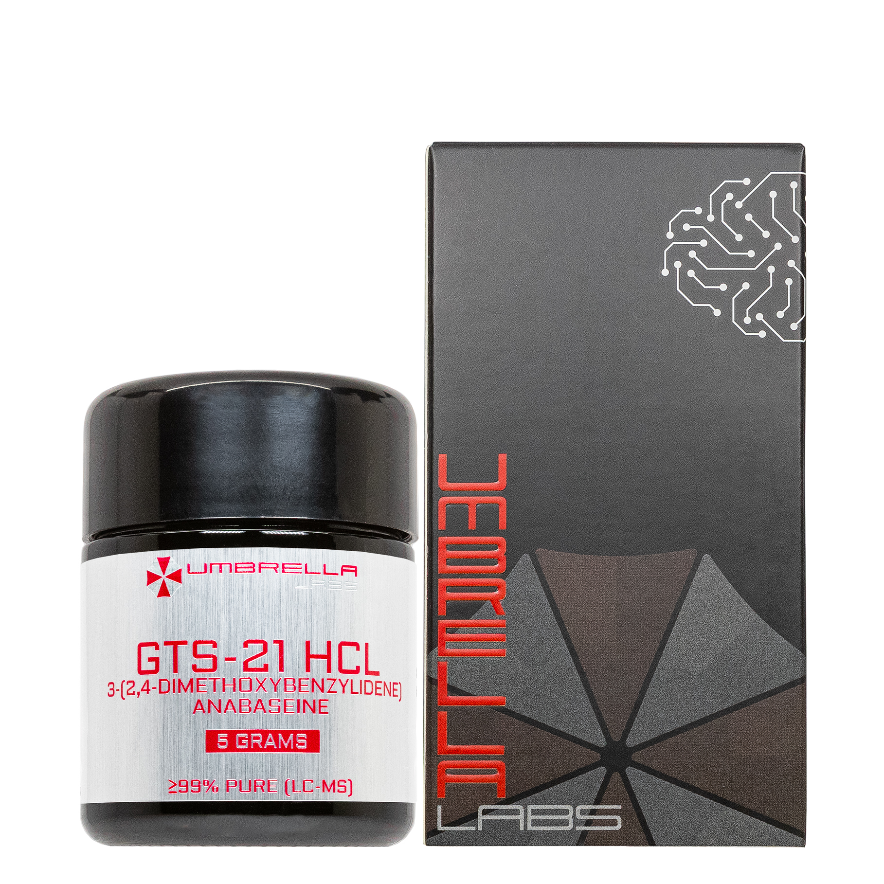
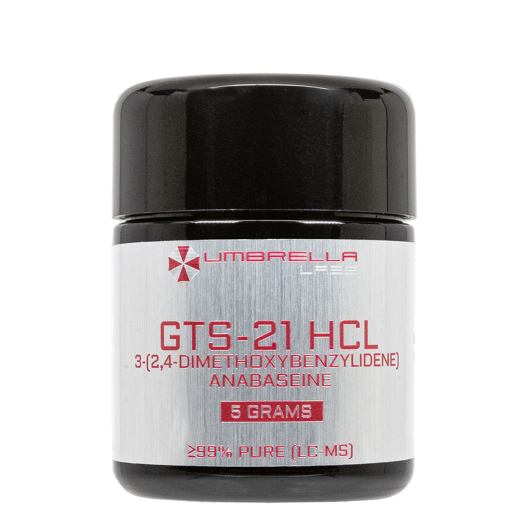


Description
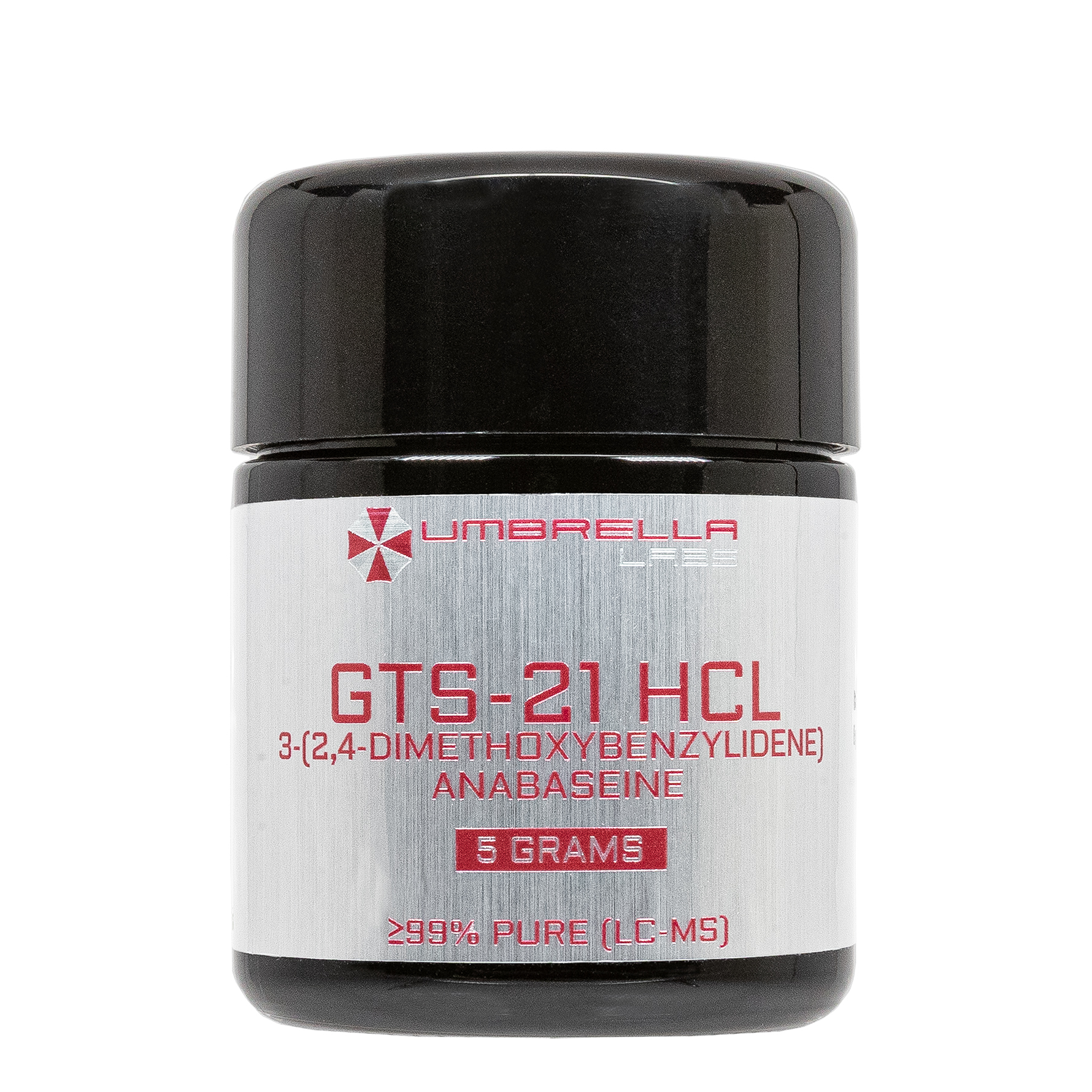
GTS-21 HCl Nootropic Powder






| CAS Number | 148372-04-7 |
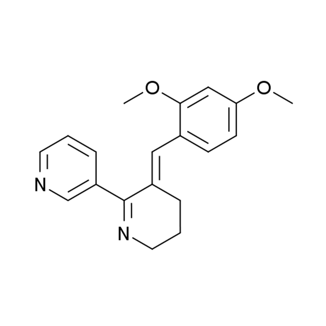
| Other Names | 3-(2,4-dimethoxy-benzylidene)anabaseine, DMXB-A, Dimethoxybenzylidene anabaseine |
| IUPAC Name | 3-[(5E)-5-[(2,4-dimethoxyphenyl)methylidene]-3,4-dihydro-2H-pyridin-6-yl]pyridine |
| Molecular Formula | C₁₉H₂₀N₂O₂ |
| Molecular Weight | 308.37 |
| Purity | ≥99% Pure (LC-MS) |
| Powder Availability |  5 grams |
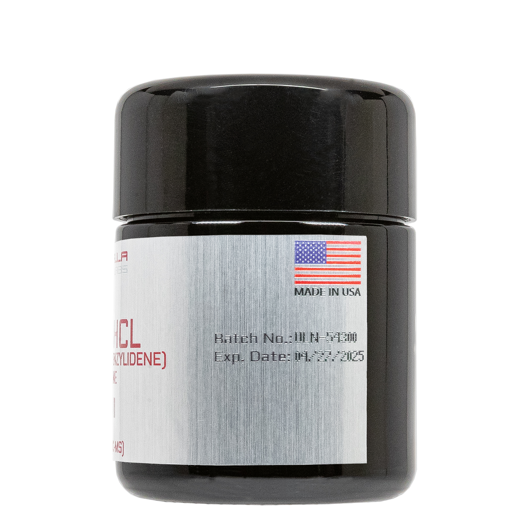
| Storage | Store in cool dry environment, away from direct sunlight. |
| Terms | All products are for laboratory developmental research USE ONLY. Products are not for human consumption. |
What is GTS-21?
GTS-21, also known as DMXBA (3-(2,4-dimethoxybenzylidene)anabaseine), is an experimental nootropic compound and selective partial agonist of the α7 nicotinic acetylcholine receptor (α7 nAChR) that has been extensively investigated for its potential to enhance cognitive function and improve neurological and psychiatric disorders. By selectively stimulating α7 nAChRs, GTS-21 enhances cholinergic neurotransmission while also indirectly modulating glutamatergic, dopaminergic, and anti-inflammatory signaling pathways involved in learning, memory, attention, and executive function. Preclinical studies have demonstrated that GTS-21 improves performance in a variety of cognitive tasks, enhances synaptic plasticity, and protects neurons against excitotoxic and inflammatory injury. These findings have prompted clinical investigations evaluating its therapeutic potential in conditions such as Alzheimer’s disease, schizophrenia, age-related cognitive decline, and inflammatory disorders, where deficits in α7 receptor signaling are believed to contribute to impaired cognition.
Main Research Findings
1) Administration of GTS-21 was shown to alleviate fibrillations related to sepsis through the modulation of macrophage polarization.
2) GTS-21 was found to reduce hepatic injury induced by polymicrobial sepsis through modulation of autophagy through α7nAChR.
Selected Data
1) The study by researchers Zhou et al was designed to investigate whether GTS-21, a selective α7nAChR agonist, could reduce the development of sepsis-induced atrial fibrillation (AF) by regulating macrophage polarization, suppressing inflammation, preserving mitochondrial function, and maintaining calcium homeostasis within the myocardium. To address this objective, the investigators combined an in vivo mouse model of lipopolysaccharide (LPS)-induced sepsis with extensive molecular, cellular, and physiological analyses. The experimental strategy incorporated survival studies, electrophysiological assessment of atrial fibrillation, echocardiography, histological examination, biochemical assays, flow cytometry, immunofluorescence, western blotting, quantitative PCR, RNA sequencing, and in vitro macrophage-cardiomyocyte co-culture experiments. Together, these complementary approaches allowed the researchers to characterize the effects of GTS-21 from the whole-animal level down to intracellular signaling pathways [1].
Eight-week-old specific pathogen-free male C57BL/6 mice were housed under standardized environmental conditions with unrestricted access to food and water following institutional animal care guidelines. Mice were randomly assigned to three experimental groups consisting of saline-treated controls (PBS), LPS-treated animals, and mice receiving GTS-21 before LPS administration. Sepsis was induced through intraperitoneal injection of lipopolysaccharide at 10 mg/kg, while GTS-21 was administered intraperitoneally at 20 mg/kg thirty minutes before LPS injection. Separate experiments evaluating survival used a higher LPS dose of 40 mg/kg to generate severe sepsis for Kaplan-Meier survival analysis over a 48-hour observation period. After treatment, animals underwent echocardiographic evaluation to assess left ventricular ejection fraction, diastolic function, and atrial structural changes before blood and cardiac tissues were collected for biochemical and histological analyses. Hearts were fixed, paraffin embedded, sectioned, and prepared for immunohistochemistry, immunofluorescence, and reactive oxygen species staining [1].
Susceptibility to atrial fibrillation was evaluated using transthoracic electrical stimulation under light isoflurane anesthesia. Surface electrocardiography monitored cardiac rhythm while electrical stimuli were delivered across the thorax to induce atrial fibrillation. AF induction was confirmed by disappearance of P waves and appearance of irregular fibrillatory waves. The investigators recorded both AF inducibility and episode duration, restoring sinus rhythm with low-energy shocks when necessary. Cardiac injury was further evaluated by measuring plasma concentrations of creatine kinase-MB (CK-MB), cardiac troponin I (cTnI), lactate dehydrogenase (LDH), and inflammatory cytokines using commercially available ELISA kits. Oxidative stress within atrial tissue was assessed using dihydroethidium staining for reactive oxygen species, while transmission electron microscopy was employed to examine mitochondrial morphology, including cristae integrity, mitochondrial fragmentation, and fusion. Additional analyses measured the expression of mitochondrial fusion protein MFN2, fission protein DRP1, calcium-handling proteins NCX-1, RYR2, and SERCA2a, as well as CaMKII phosphorylation using western blotting and RT-qPCR [1].
To investigate immune mechanisms, primary peritoneal macrophages were isolated from thioglycolate-treated mice and cultured under inflammatory conditions. Macrophages were pretreated with GTS-21 before stimulation with LPS for twenty-four hours. Flow cytometry quantified macrophage polarization using CD86 and inducible nitric oxide synthase (iNOS) as M1 markers and CD206 and arginase-1 (Arg-1) as M2 markers. Western blotting, RT-qPCR, immunofluorescence, and ELISA were used to evaluate inflammatory signaling pathways, including NF-κB P65 activation, NLRP3 inflammasome assembly, caspase-1 cleavage, and production of inflammatory cytokines such as IL-1β, IL-6, and IL-18. Nuclear translocation of NF-κB P65 was visualized by confocal microscopy to determine whether GTS-21 inhibited inflammatory transcriptional activation within macrophages.
The investigators next established an indirect macrophage-cardiomyocyte co-culture model using HL-1 atrial cardiomyocytes. Conditioned media collected from macrophages treated with PBS, LPS, or LPS plus GTS-21 were mixed with HL-1 culture medium to determine how macrophage-derived inflammatory factors influenced cardiomyocyte function. Reactive oxygen species production, mitochondrial membrane potential, intracellular calcium concentration, mitochondrial fusion and fission proteins, and calcium-regulating proteins were measured using confocal microscopy, flow cytometry, JC-1 fluorescence staining, western blotting, and RT-qPCR. Finally, transcriptomic analysis of atrial tissue was performed using high-throughput RNA sequencing. Differentially expressed genes were identified with DESeq2 and integrated with secreted protein databases and protein-protein interaction network analyses to identify candidate mediators of GTS-21’s cardioprotective effects. Neuregulin-1 (NRG1) emerged as a prominent candidate, and its expression and secretion were subsequently validated using RT-qPCR, western blotting, and ELISA. Neutralizing antibodies against NRG1 were then added to conditioned media to determine whether macrophage-derived NRG1 mediated the beneficial effects of GTS-21 on oxidative stress, mitochondrial integrity, and calcium homeostasis in cardiomyocytes [1].
2) The study performed by Wu et al was designed to determine whether activation of the α7nAChR by the selective agonist GTS-21 could protect against polymicrobial sepsis-induced liver injury by suppressing inflammation and enhancing autophagy. To investigate this hypothesis, the researchers employed a murine cecal ligation and puncture (CLP) model of polymicrobial sepsis and combined histological, biochemical, ultrastructural, and molecular analyses to evaluate hepatic injury, inflammatory cytokine production, and autophagy-related signaling. By comparing septic animals treated with GTS-21 to untreated septic controls and animals receiving the α7nAChR antagonist α-bungarotoxin (α-BGT), the investigators sought to determine whether the protective effects of GTS-21 were mediated specifically through activation of α7 nicotinic acetylcholine receptors [2].
Forty male C57BL/6 mice aged 8–10 weeks were housed under specific pathogen-free conditions with free access to food and water, and all procedures were approved by the institutional animal care committee. Polymicrobial sepsis was induced using the established CLP model. Animals were anesthetized with intraperitoneal ketamine and midazolam before a midline laparotomy was performed. The cecum was exteriorized, ligated at approximately 60% of its length using silk suture, and punctured twice with a 21-gauge needle to permit leakage of intestinal contents into the peritoneal cavity. After the cecum was returned to the abdomen, the incision was closed and mice received subcutaneous normal saline resuscitation. Sham-operated mice underwent identical surgical procedures except for cecal ligation and puncture. This model was selected because it closely reproduces the systemic inflammatory response and multiple-organ injury observed during clinical polymicrobial sepsis [2].
Animals were randomly assigned to one of four experimental groups containing ten mice each: a sham-operated control group, a CLP group, a CLP plus GTS-21 treatment group, and an α-BGT plus CLP plus GTS-21 group. Mice assigned to the treatment group received intraperitoneal GTS-21 at a dose of 4.0 mg/kg immediately after CLP surgery. To determine whether the effects of GTS-21 required activation of α7nAChRs, animals in the antagonist group received a 1.0 μg/kg intraperitoneal dose of α-bungarotoxin before CLP, followed by GTS-21 administration after surgery. Six hours after the procedure, blood samples and liver tissues were collected for biochemical, histological, ultrastructural, and molecular analyses. This relatively early time point was selected to evaluate acute inflammatory responses and early alterations in autophagy during sepsis.
To assess hepatic injury, liver specimens were fixed in formaldehyde, embedded in paraffin, sectioned into 4-μm slices, and stained with hematoxylin and eosin (H&E). Histological damage was evaluated microscopically using the Knodell Histology Activity Index, which grades hepatic necrosis and inflammatory infiltration. Plasma alanine aminotransferase (ALT) and aspartate aminotransferase (AST) concentrations were measured as biochemical indicators of liver injury using an automated chemistry analyzer. Systemic inflammation was assessed by measuring plasma tumor necrosis factor-α (TNF-α) and interleukin-6 (IL-6) concentrations with enzyme-linked immunosorbent assay (ELISA). Together, these measurements provided quantitative assessments of hepatic damage and inflammatory activity following septic challenge [2].
The investigators next evaluated autophagy using both ultrastructural and molecular approaches. Transmission electron microscopy (TEM) was performed on glutaraldehyde-fixed liver tissue to directly visualize autophagic vacuoles and quantify autophagosome formation. Liver samples were processed through osmium tetroxide fixation, dehydration, resin embedding, ultrathin sectioning, and heavy-metal staining before examination with a transmission electron microscope. In parallel, western blot analysis was performed on liver homogenates to measure proteins involved in autophagy. Equal amounts of protein were separated by SDS-PAGE, transferred to polyvinylidene fluoride membranes, and probed with antibodies against LC3-II, p62/SQSTM1, Atg5, Atg7, and GAPDH. LC3-II served as a marker of autophagosome formation, p62 accumulation was used to evaluate autophagic flux, and Atg5 and Atg7 were examined because of their essential roles in autophagosome elongation and maturation. Band intensities were normalized to GAPDH before quantitative analysis.
Finally, statistical analyses were performed using SPSS software. All data was expressed as the mean ± standard error. Comparisons among the four experimental groups were conducted using one-way ANOVA followed by Bonferroni post hoc testing to identify significant pairwise differences. A probability value below 0.05 was considered statistically significant. By integrating pathological examination, serum biomarkers, ultrastructural imaging, inflammatory cytokine measurements, and analyses of autophagy-associated proteins, the investigators comprehensively evaluated whether activation of α7 nicotinic acetylcholine receptors by GTS-21 could alleviate sepsis-induced hepatic injury through regulation of inflammatory responses and restoration of autophagic activity [2].
Discussion
1) The study conducted by researchers Zhou et al demonstrated that GTS-21 significantly protected against sepsis-induced AF by improving survival, reducing myocardial injury, suppressing inflammatory macrophage activation, preserving mitochondrial function, restoring calcium homeostasis, and enhancing the secretion of the cardioprotective factor NRG1. In mice challenged with LPS, severe systemic inflammation markedly increased susceptibility to AF while simultaneously impairing cardiac function and survival. Administration of GTS-21 substantially improved these outcomes. In the severe sepsis model using 40 mg/kg LPS, mice treated with GTS-21 exhibited significantly higher 48-hour survival rates than untreated septic animals. Using the lower-dose LPS model to evaluate arrhythmias, GTS-21 significantly reduced both the incidence and duration of electrically induced AF [1].
Echocardiographic measurements further demonstrated that GTS-21 preserved left ventricular systolic performance by restoring left ventricular ejection fraction and significantly improved diastolic function, as reflected by normalization of the E/A and E’/A’ ratios. Circulating biomarkers of myocardial injury, including CK-MB, cTnI, and LDH, were all significantly elevated in septic mice but were markedly reduced following GTS-21 treatment, indicating substantial protection against inflammation-induced cardiac damage.
The investigators next examined whether these physiological improvements were associated with changes in cardiac inflammation and macrophage behavior. Immunofluorescence staining demonstrated a pronounced accumulation of F4/80-positive macrophages within atrial tissue after LPS administration, whereas GTS-21 markedly reduced macrophage infiltration. Molecular analyses revealed that septic mice exhibited a strong shift toward the pro-inflammatory M1 macrophage phenotype, characterized by increased expression of M1 markers and elevated inflammatory cytokines including IL-1β, IL-6, and IL-18. Treatment with GTS-21 reversed these changes by suppressing M1 polarization while promoting differentiation toward the anti-inflammatory M2 phenotype. These findings indicated that activation of α7 nicotinic acetylcholine receptors effectively altered the inflammatory environment within atrial tissue, thereby reducing cytokine production and limiting inflammation-driven atrial remodeling that contributes to arrhythmogenesis [1].
The study also demonstrated that GTS-21 protected mitochondrial integrity and reduced oxidative stress within cardiac tissue. LPS-induced sepsis produced marked mitochondrial abnormalities, including disrupted mitochondrial organization, fragmented cristae, excessive mitochondrial fission, and reduced mitochondrial fusion. Electron microscopy showed that GTS-21 substantially restored normal mitochondrial morphology. These structural improvements were supported by molecular findings showing decreased expression of the fission protein DRP1 and increased expression of the fusion protein MFN2 after treatment. Consistent with improved mitochondrial health, GTS-21 significantly reduced reactive oxygen species (ROS) accumulation within atrial tissue. Because oxidative stress contributes directly to calcium dysregulation during atrial fibrillation, the investigators measured calcium-handling proteins and found that septic mice exhibited elevated NCX-1, RYR2, CaMKII, and phosphorylated CaMKII together with reduced SERCA2a expression. GTS-21 reversed these abnormalities, indicating restoration of intracellular calcium handling and prevention of calcium overload, both of which reduce susceptibility to atrial fibrillation.
Experiments using primary peritoneal macrophages further clarified the molecular mechanisms responsible for these protective effects. LPS stimulation strongly promoted M1 macrophage polarization, increased expression of CD86 and inducible nitric oxide synthase (iNOS), activated the NF-κB P65/NLRP3/caspase-1 signaling pathway, and stimulated production of inflammatory cytokines. GTS-21 significantly inhibited each of these inflammatory responses. Flow cytometry confirmed reduced M1 polarization and enhanced M2 polarization, while western blotting and RT-qPCR demonstrated suppression of NF-κB P65, NLRP3, and cleaved caspase-1 expression. Confocal microscopy showed that GTS-21 prevented nuclear translocation of NF-κB P65, thereby limiting activation of downstream inflammatory pathways. Consequently, secretion of IL-1β, IL-6, and IL-18 was significantly reduced, providing strong evidence that GTS-21 interrupts macrophage-driven inflammatory signaling at multiple levels [1].
Using an indirect co-culture system, the investigators demonstrated that conditioned media from LPS-stimulated macrophages induced substantial oxidative stress, mitochondrial dysfunction, and intracellular calcium overload in HL-1 cardiomyocytes. In contrast, conditioned media from macrophages treated with both LPS and GTS-21 significantly reduced ROS accumulation, normalized mitochondrial membrane potential, restored the balance between mitochondrial fusion and fission, and corrected calcium-handling protein expression. RNA sequencing identified NRG1 as one of the most significantly upregulated secreted proteins following GTS-21 treatment. Subsequent RT-qPCR, western blotting, and ELISA confirmed increased NRG1 expression and secretion by macrophages. Importantly, neutralization of NRG1 largely abolished the beneficial effects of GTS-21, resulting in renewed oxidative stress, mitochondrial injury, intracellular calcium overload, and dysregulation of NCX-1, RYR2, SERCA2a, and phosphorylated CaMKII in cardiomyocytes.These findings demonstrated that macrophage-derived NRG1 serves as a critical downstream mediator of GTS-21-induced cardioprotection [1].
Overall, the results establish that GTS-21 reduces sepsis-induced atrial fibrillation by promoting anti-inflammatory macrophage polarization, suppressing NF-κB/NLRP3 inflammasome activation, enhancing NRG1 secretion, preserving mitochondrial function, reducing oxidative stress, restoring calcium homeostasis, and ultimately protecting the myocardium from inflammation-induced electrical and structural remodeling [1].
2) The study performed by the research team of Wu et al demonstrated that activation of α7 nicotinic acetylcholine receptors with GTS-21 significantly reduced polymicrobial sepsis-induced hepatic injury by suppressing inflammatory responses and enhancing autophagy. Histological examination of liver tissue revealed that sham-operated mice exhibited normal hepatic architecture with well-organized liver sinusoids and healthy hepatocytes. In contrast, mice subjected to CLP developed severe hepatic injury characterized by extensive mononuclear inflammatory cell infiltration, sinusoidal congestion, hepatocyte swelling, necrosis, and formation of localized abscesses surrounding damaged liver cells. These pathological changes were accompanied by significantly elevated histological necrosis scores [2].
Administration of GTS-21 immediately after CLP markedly improved liver morphology, preserving hepatic strand organization, reducing inflammatory infiltration, limiting hepatocyte degeneration, and significantly lowering liver injury scores compared with untreated septic animals. However, pretreatment with the α7 nicotinic acetylcholine receptor antagonist α-bungarotoxin (α-BGT) largely abolished these protective effects, resulting in persistent hepatic edema, eosinophilic degeneration, inflammatory infiltration, and histological damage comparable to that observed in untreated CLP mice. These findings demonstrated that activation of α7nAChRs is essential for the hepatoprotective effects produced by GTS-21 during polymicrobial sepsis.
Biochemical analyses supported the histological observations by demonstrating significant reductions in both hepatic injury markers and systemic inflammatory cytokines following GTS-21 treatment. CLP-induced sepsis caused marked elevations in plasma ALT and AST, indicating substantial hepatocellular damage. Compared with sham animals, ALT and AST values increased several-fold following CLP. Treatment with GTS-21 significantly lowered both enzymes, indicating preservation of hepatocyte integrity and improved liver function. Similarly, systemic inflammation was markedly increased after CLP, as reflected by elevated circulating concentrations of TNF-α and IL-6. GTS-21 significantly reduced both pro-inflammatory cytokines, demonstrating that activation of the cholinergic anti-inflammatory pathway effectively suppressed the excessive inflammatory response associated with sepsis. Once again, administration of α-BGT reversed these beneficial effects, restoring high concentrations of ALT, AST, TNF-α, and IL-6. These biochemical findings confirmed that the anti-inflammatory and hepatoprotective actions of GTS-21 were dependent upon α7 nicotinic acetylcholine receptor activation rather than nonspecific pharmacological effects [2].
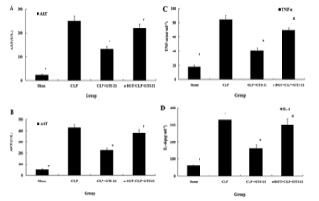
Figure 1: Changes in levels of hepatic enzymes and plasma pro-inflammatory cytokines
Transmission electron microscopy provided additional evidence that GTS-21 enhanced hepatic autophagy during sepsis. Ultrastructural examination showed relatively few autophagic vacuoles in sham-operated mice. CLP-induced sepsis significantly increased the number of autophagosomes, indicating activation of the autophagic process in response to severe cellular stress. However, GTS-21 treatment produced an even greater increase in autophagic vacuole formation than CLP alone, suggesting that activation of α7nAChRs further stimulated autophagy beyond the endogenous response to sepsis. Administration of α-BGT significantly reduced the number of autophagosomes compared with GTS-21-treated animals, indicating that the enhanced autophagic activity required α7 receptor signaling. The authors interpreted these findings as evidence that GTS-21 promotes autophagic flux, thereby facilitating removal of damaged cellular components generated during septic injury and contributing to improved hepatic preservation.
Western blot analyses further characterized changes in autophagy-related proteins. Sepsis induced by CLP significantly increased expression of LC3-II, p62, Atg5, and Atg7 compared with sham controls. Although elevated LC3-II reflected increased autophagosome formation, simultaneous accumulation of p62 suggested that autophagic degradation was impaired, resulting in incomplete autophagic flux. Treatment with GTS-21 produced a distinct pattern of protein expression that indicated restoration of functional autophagy. LC3-II expression increased further following GTS-21 administration, demonstrating enhanced autophagosome formation, while expression of the autophagy-related proteins Atg5 and Atg7 was also significantly elevated, supporting greater phagophore elongation and maturation of autophagosomes. In contrast, p62 expression was markedly reduced after GTS-21 treatment. Because p62 is normally degraded during successful autophagy, its reduction suggested that GTS-21 improved completion of the autophagic process rather than merely increasing autophagosome accumulation. When α-BGT was administered before GTS-21, these molecular changes were largely abolished. LC3-II, Atg5, and Atg7 expression decreased, whereas p62 again accumulated, demonstrating that blockade of α7 nicotinic receptors prevented restoration of normal autophagic flux [2].
Collectively, the results demonstrated that GTS-21 exerts a multifaceted protective effect during polymicrobial sepsis by simultaneously suppressing inflammation and improving hepatic autophagy through activation of α7 nicotinic acetylcholine receptors. Activation of the cholinergic anti-inflammatory pathway reduced circulating TNF-α and IL-6, limited hepatocellular injury, preserved liver architecture, and lowered serum ALT and AST concentrations. At the cellular level, GTS-21 enhanced autophagic activity by increasing LC3-II, Atg5, and Atg7 while reducing accumulation of the autophagy substrate p62, indicating restoration of autophagic flux rather than simple accumulation of autophagosomes. The consistent reversal of these beneficial effects by α-bungarotoxin established that α7nAChR activation was required for both the anti-inflammatory and pro-autophagic actions of GTS-21. Overall, the findings support the conclusion that stimulation of α7 nicotinic acetylcholine receptors protects against sepsis-induced hepatic injury by coordinating suppression of inflammatory signaling with enhancement of autophagy, thereby improving cellular homeostasis and limiting liver damage during the early stages of polymicrobial sepsis [2].
Disclaimer
**LAB USE ONLY**
*This information is for educational purposes only and does not constitute medical advice. THE PRODUCTS DESCRIBED HEREIN ARE FOR RESEARCH USE ONLY. All clinical research must be conducted with oversight from the appropriate Institutional Review Board (IRB). All preclinical research must be conducted with oversight from the appropriate Institutional Animal Care and Use Committee (IACUC) following the guidelines of the Animal Welfare Act (AWA).
Citations
[1] Zhou J, Wu K, Ma Y, et al. GTS-21 alleviates sepsis-induced atrial fibrillation susceptibility by modulating macrophage polarization and Neuregulin-1 secretion. Int Immunopharmacol. 2025;154:114561. doi:10.1016/j.intimp.2025.114561
[2] Wu XJ, Yan XT, Yang XM, et al. GTS-21 ameliorates polymicrobial sepsis-induced hepatic injury by modulating autophagy through α7nAchRs in mice. Cytokine. 2020;128:155019. doi:10.1016/j.cyto.2020.155019
GTS-21 HCl is sold for laboratory research use only. Terms of sale apply. Not for human consumption, nor medical, veterinary, or household uses. Please familiarize yourself with our Terms & Conditions prior to ordering.
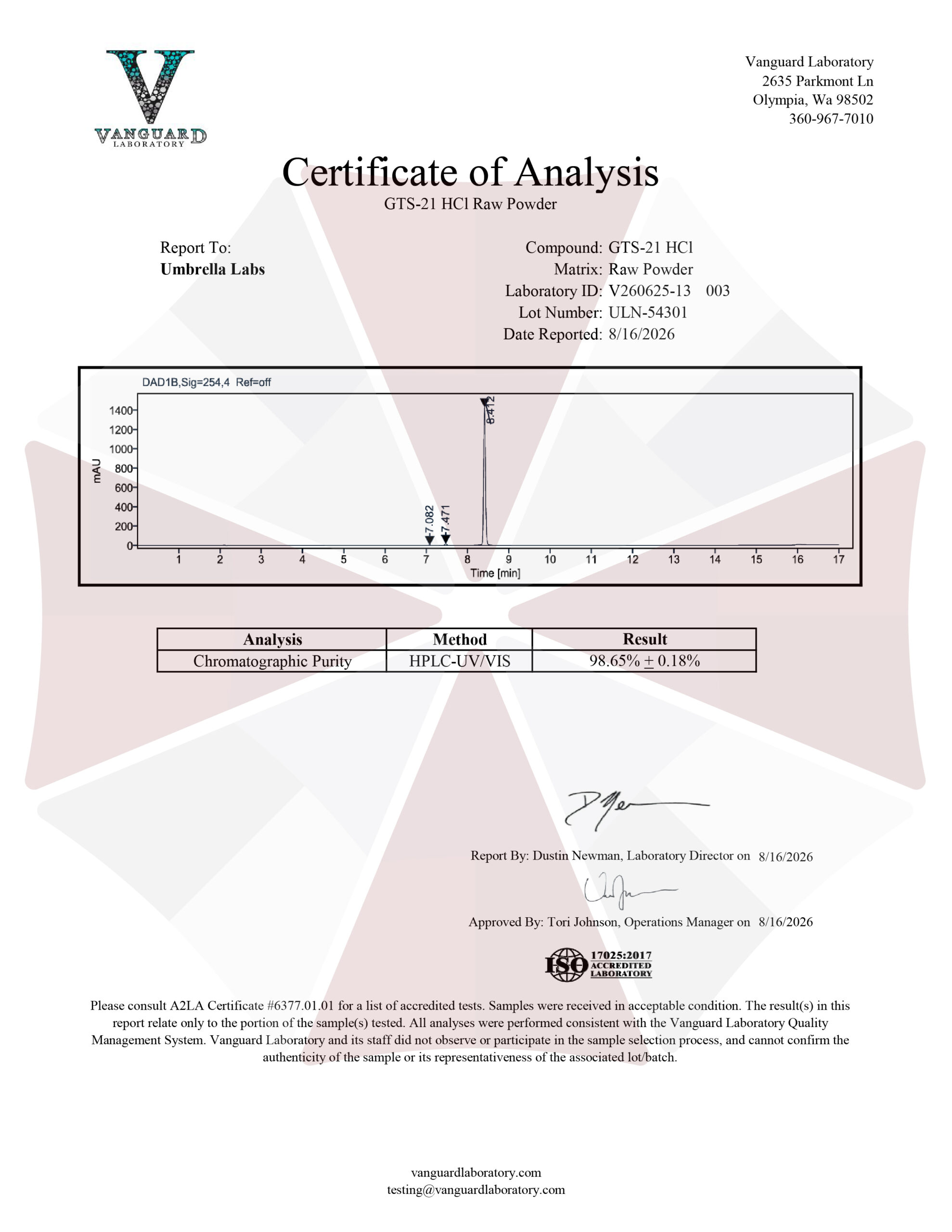
| File Name | View/Download |
| 2026-08-16-UmbrellaLabs-GTS-21-HCl-Certificate-of-Analysis-COA.pdf |  |