

Description
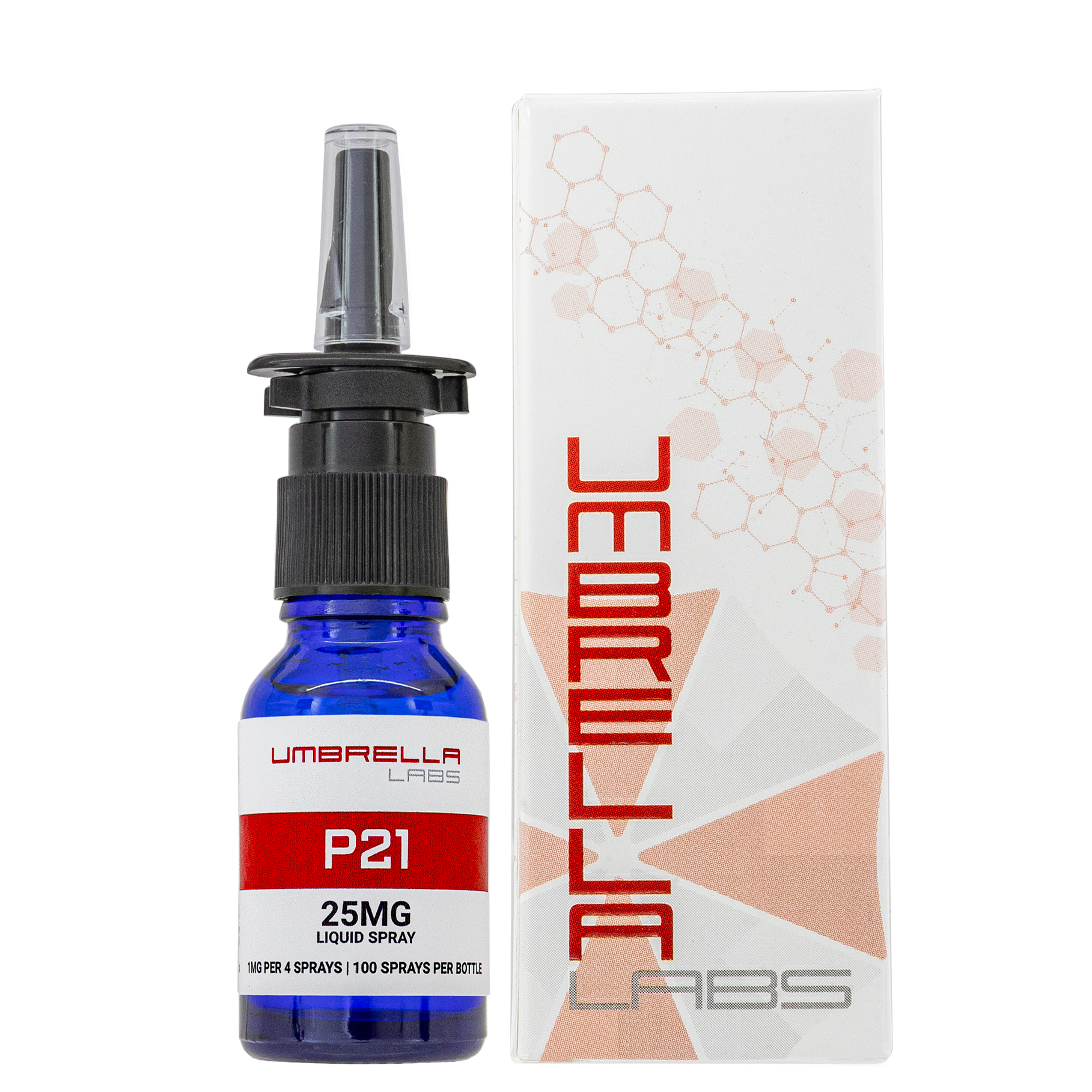
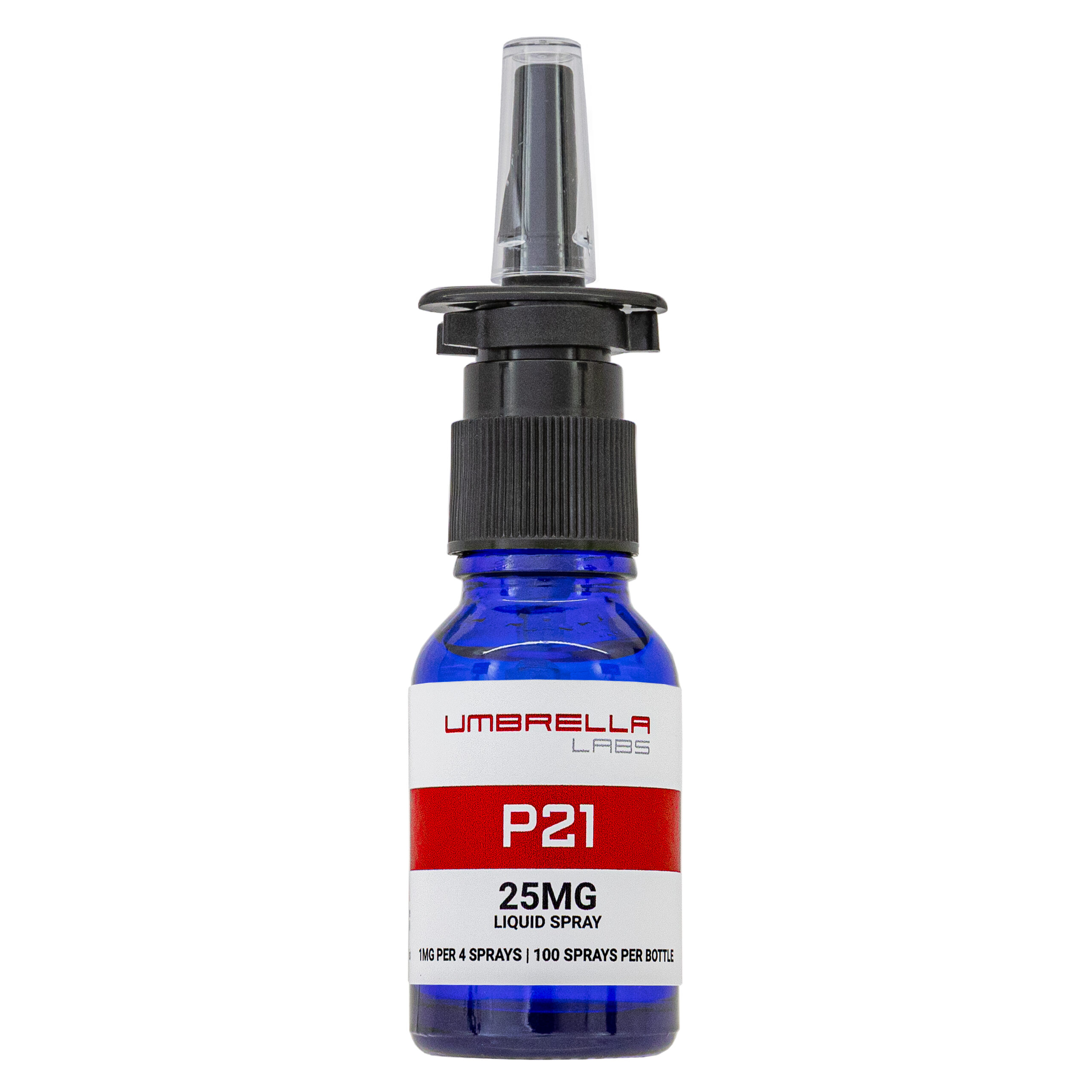
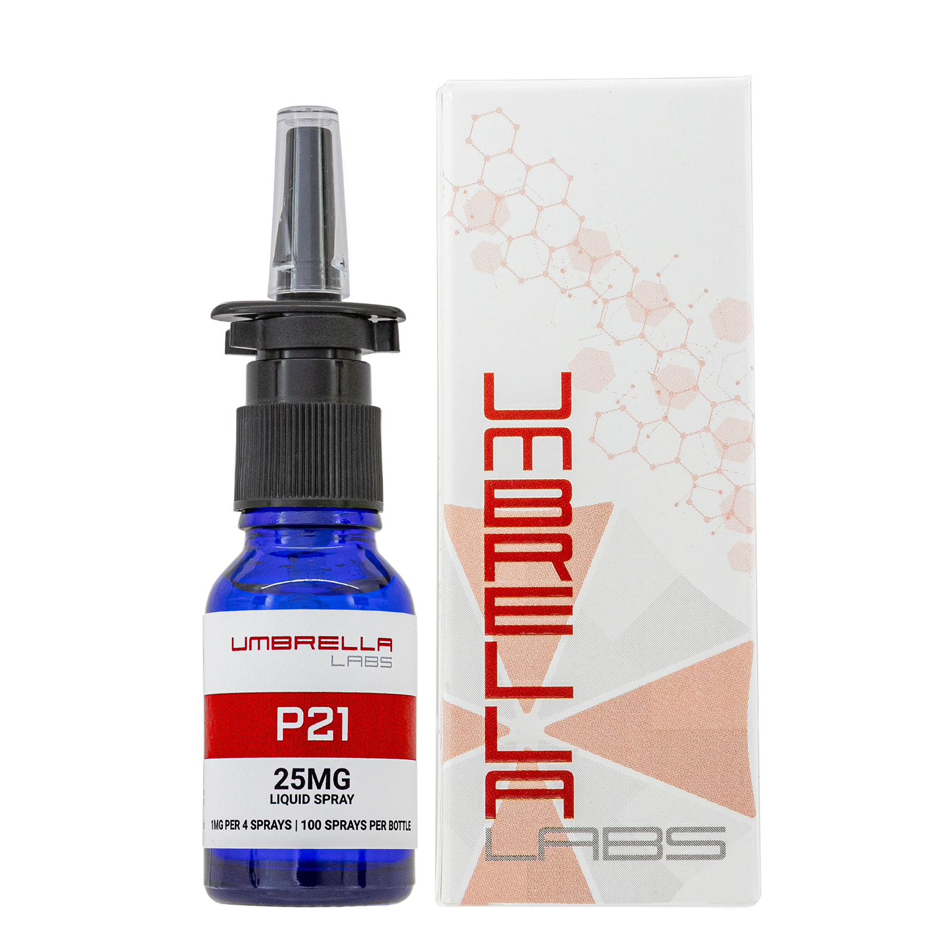
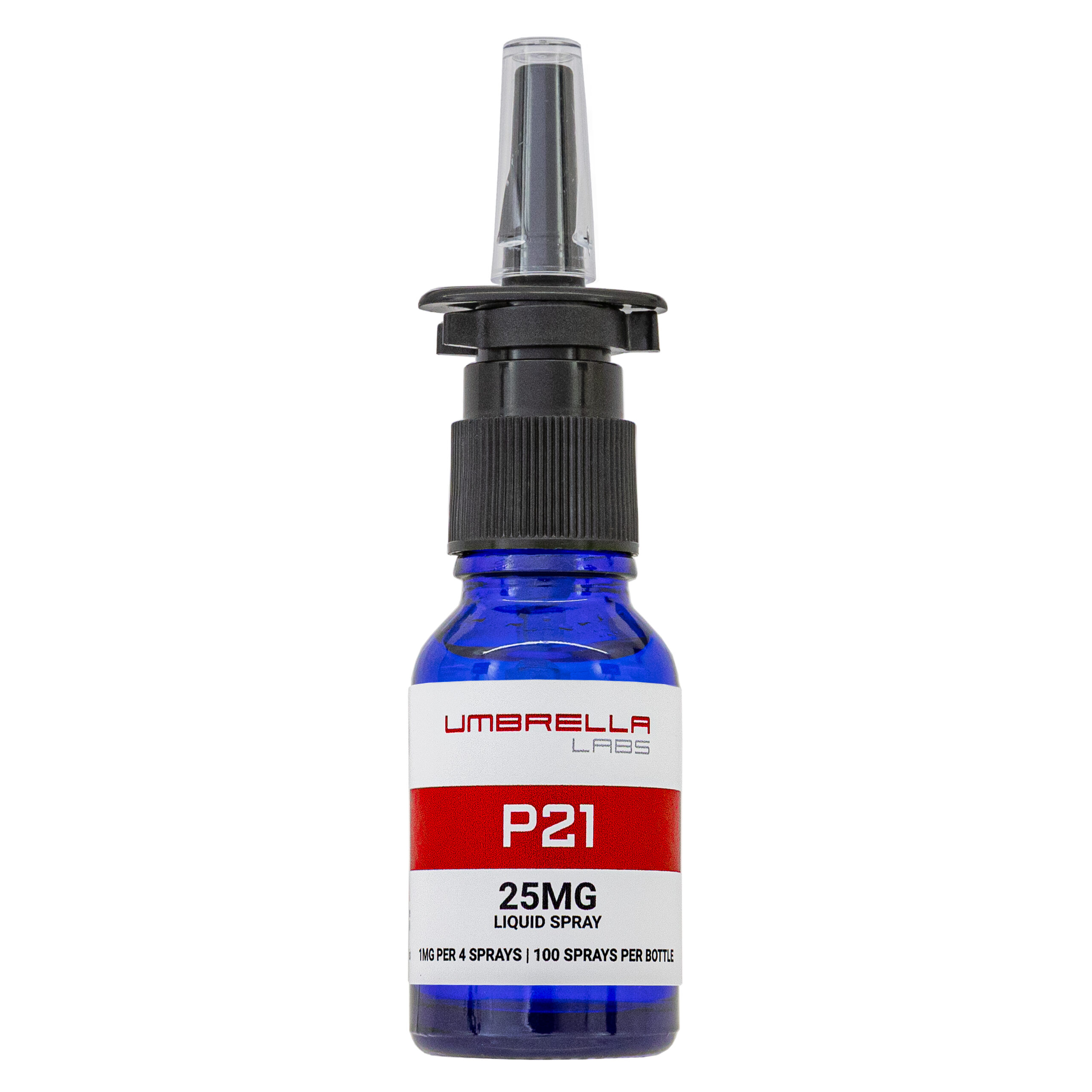
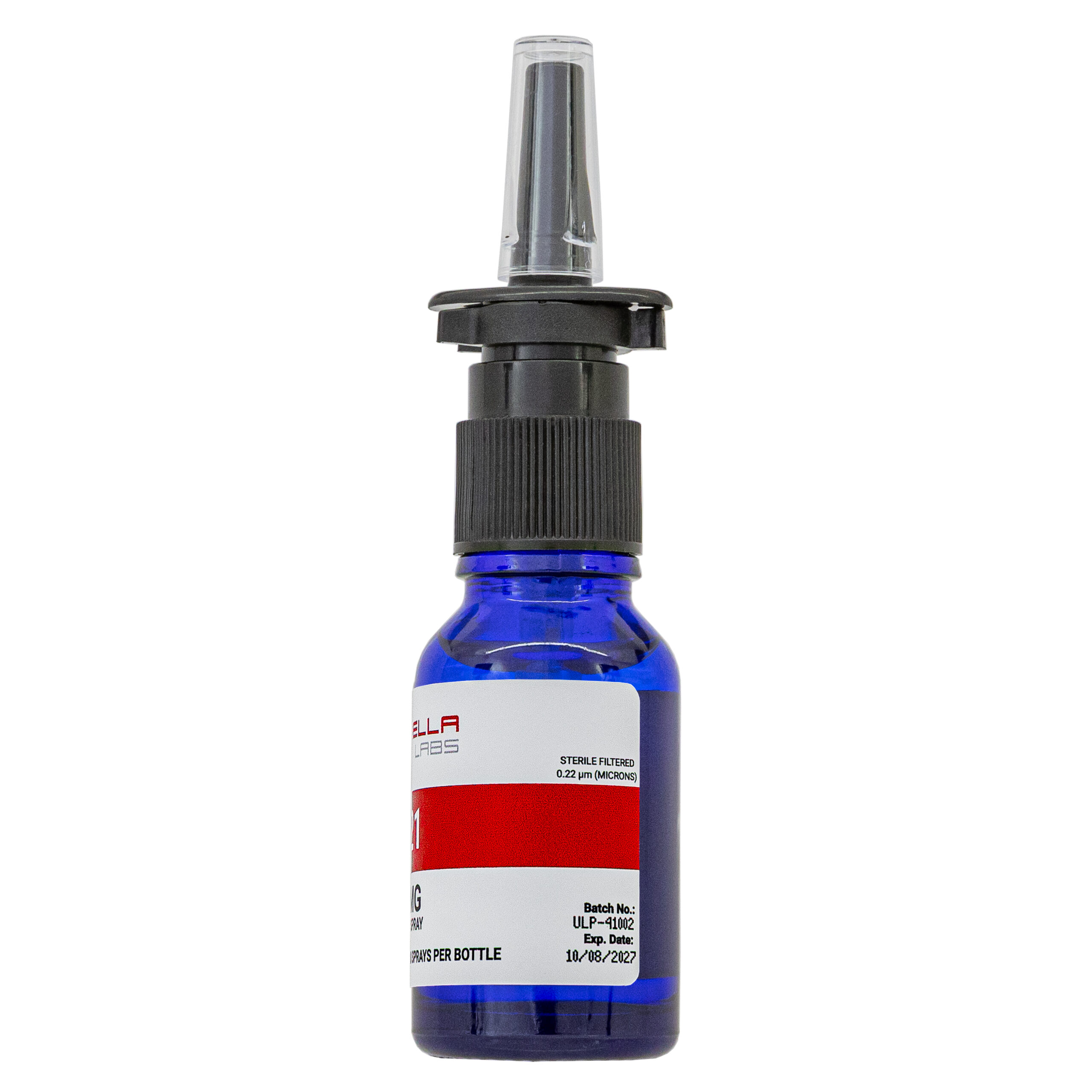
P21 Peptide Liquid Spray






| CAS Number | 1246751-68-7 |
| Other Names | P021, Peptide 021 |
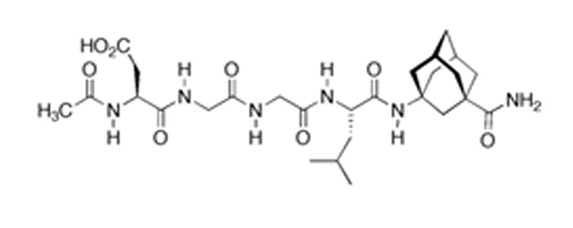
| Sequence | Ac-DGGLAG-NH2 |
| Molecular Formula | C₂₇H₄₂N₆O₈ |
| Molecular Weight | 578.66 |
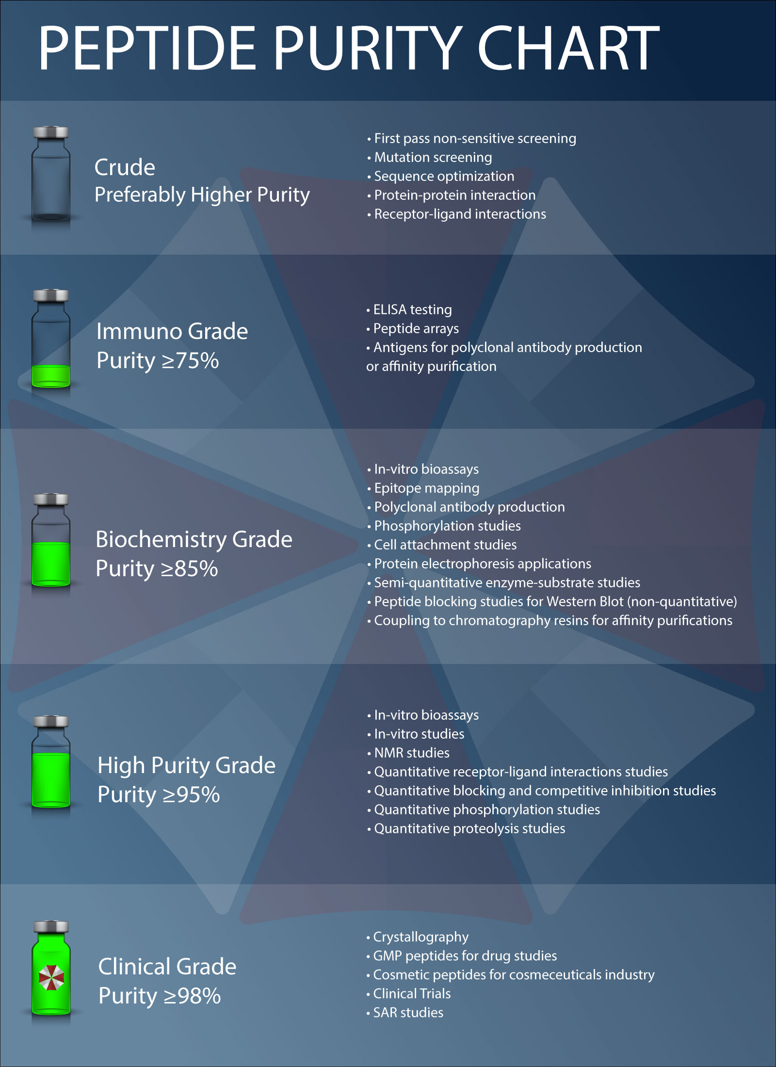
| Purity | ≥99% Pure (LC-MS) |
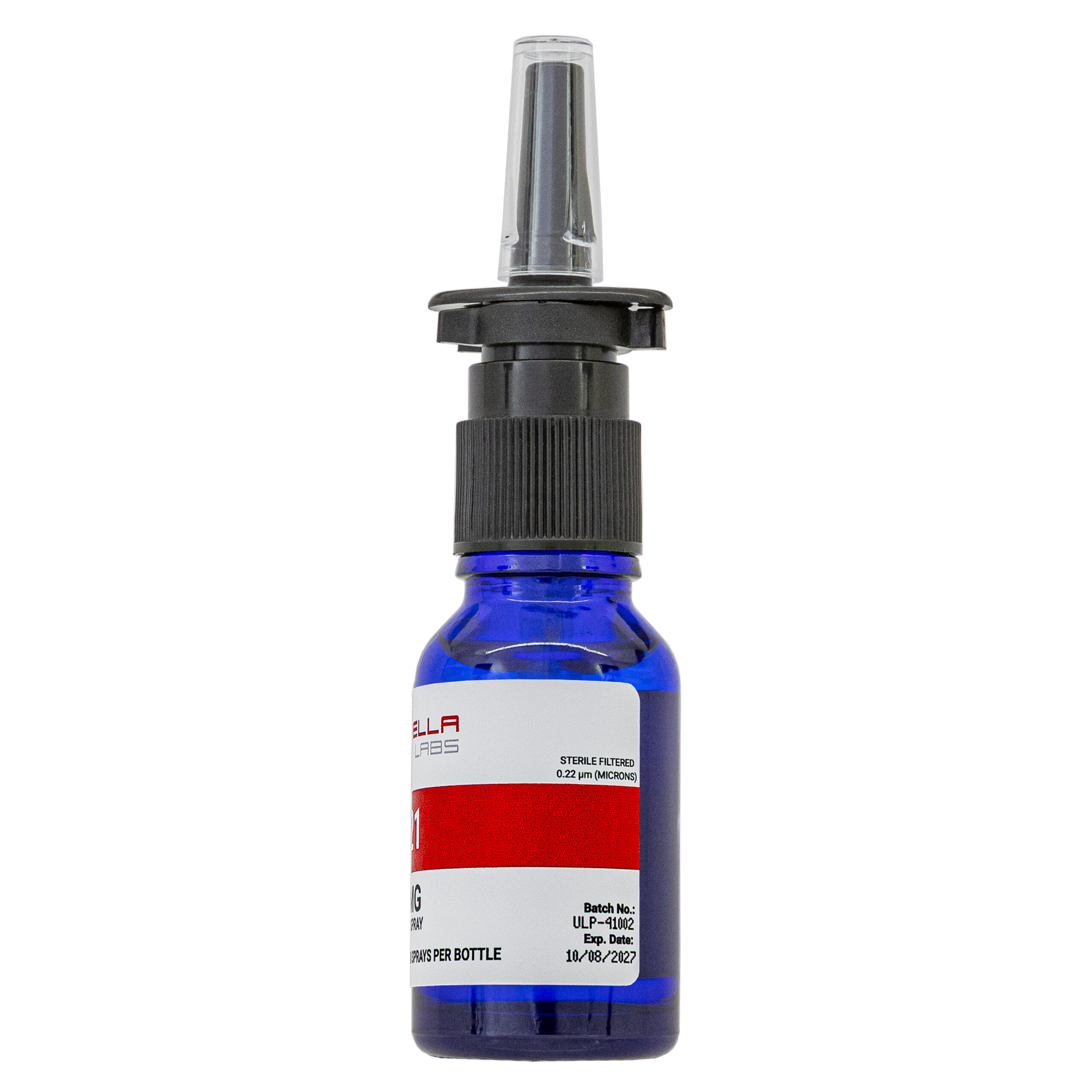
| Liquid Availability |  15mL liquid solution (250mcg/spray, 25mg total) |
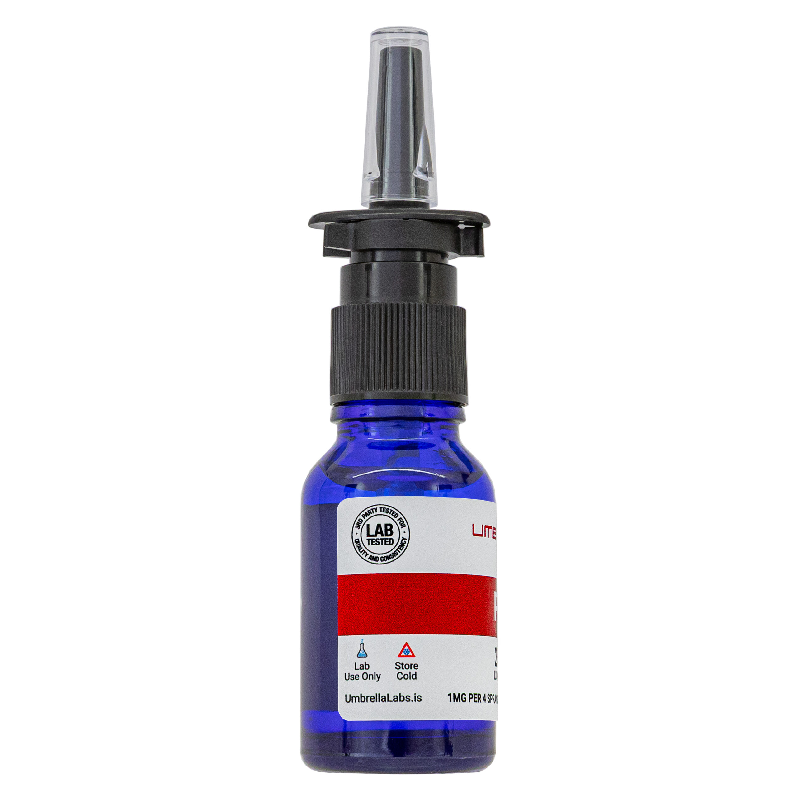
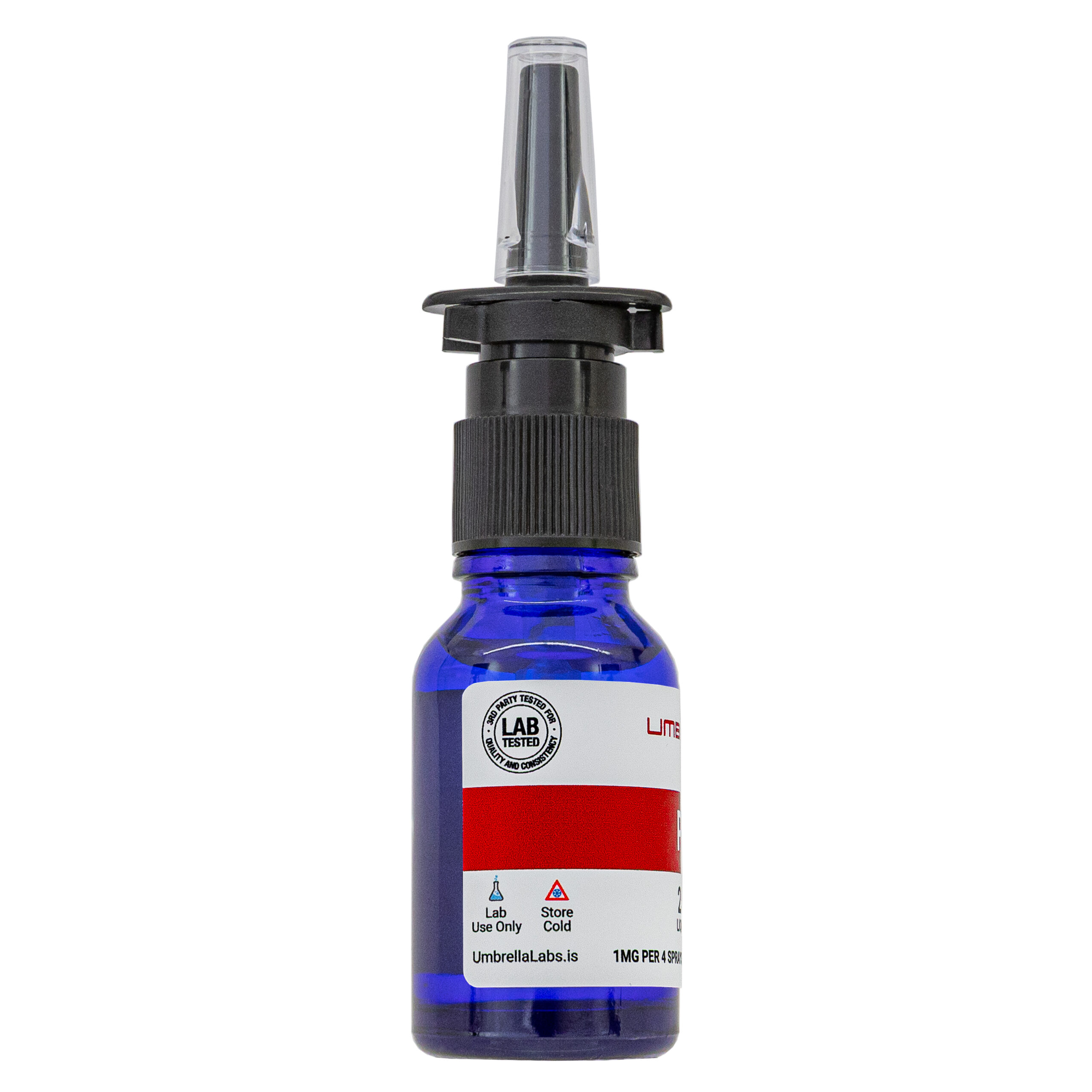
| Storage Condition | Store cold, keep refrigerated. Do NOT freeze. |
| Terms | All products are for laboratory developmental research USE ONLY. Products are not for human consumption. |
What is P21?
P21, also referred to as P021 or Peptide 021, is a small-molecule peptide mimetic derived from a biologically active region of ciliary neurotrophic factor (CNTF) that has been investigated for its potential neuroregenerative and cognitive-enhancing properties. P021 was developed by narrowing the larger CNTF-derived Peptide 6 to its minimal active sequence and subsequently modifying the structure with an adamantylated glycine group to improve its ability to cross the blood-brain barrier. The compound has demonstrated neurogenic and neurotrophic activity in preclinical models. Research summarized in the article indicates that P021 can enhance adult hippocampal progenitor proliferation and differentiation, increase neurogenesis and synaptic marker expression, and improve cognitive performance. In aged and Alzheimer’s disease models, P021 has also been associated with increased brain-derived neurotrophic factor (BDNF) expression, reduced abnormal tau phosphorylation, improved hippocampal neurogenesis, and amelioration of dendritic and synaptic deficits.
Main Research Findings
1) P021 was shown to improve synaptic and dendritic deficits, increase neurogenesis, and reduce cognitive impairments in a mouse model of Alzheimer’s disease.
2) The review of the compound P021 reported on the safety, pharmacokinetics, and potential therapeutic benefits of the compound for the treatment of Alzheimer’s disease.
Selected Data
1) The study completed by the research team of Baazaoui and Iqbal investigated whether chronic administration of Peptide 021 (P021/P21), a synthetic neurotrophic/neurogenic peptide derived from the active region of ciliary neurotrophic factor (CNTF), could prevent dendritic and synaptic deficits, enhance neurogenesis, and preserve cognitive function in a transgenic mouse model of Alzheimer’s disease (AD). The investigators used female triple-transgenic AD (3 × Tg-AD) mice, a model expressing the human APPSwe mutation, tau P301L mutation, and a presenilin-1 (PS1) knock-in on a 129/Sv × C57BL/6 genetic background. Age-matched wild-type (WT) mice from the same genetic background served as controls. The animals were housed under standardized laboratory conditions, with five mice per cage, a 12-hour light/dark cycle, and unrestricted access to food and water. Only female mice were used because previous work indicated that female 3 × Tg-AD mice develop more consistent and aggressive amyloid pathology, earlier cognitive impairment, and more pronounced deficits in neurogenesis than males. Behavioral studies and neurobiological analyses were performed using separate groups of animals so that behavioral testing itself would not influence measurements of neurogenesis or synaptic plasticity [1].
The experimental design consisted of 41 WT mice, 35 vehicle-treated 3 × Tg-AD mice, and 32 P021-treated 3 × Tg-AD mice. Beginning at approximately three months of age, the P021 group received chow containing 60 nmol of P021 per gram of feed, whereas both the transgenic vehicle group and WT controls received identical chow without P021. The animals consumed approximately 2.7 g of feed per day, corresponding to an estimated intake of approximately 162 nmol of P021 per mouse per day. Food consumption was recorded every two weeks and body weight was measured monthly. P021 itself was described as a pentapeptide, Ac-DGGLAG-NH2, with a molecular weight of 578.3 Da. It was generated by adding adamantylated glycine to the C-terminal end of a CNTF-derived tetrapeptide. This modification was intended to improve blood-brain barrier permeability and reduce degradation by carboxypeptidases. The compound was synthesized and purified using reverse-phase HPLC to greater than 96% purity, with its sequence confirmed by mass spectrometry. The investigators also reported that P021 demonstrated greater than 95% stability in artificial gastric juice for 30 minutes, approximately 100% stability in intestinal fluid for 120 minutes, and plasma stability exceeding three hours, supporting its use through dietary administration [1].
The study evaluated the animals at several time points to determine both early and long-term effects of P021. Brain samples from animals that did not undergo behavioral testing were collected at three months, immediately before treatment began, and again at nine, 15, and 18 months after treatment. Behavioral testing was conducted after nine months of treatment using the Morris Water Maze and after approximately 15–16 months of treatment using a novel object location paradigm. The Morris Water Maze was used to evaluate spatial reference memory. Mice were trained in a 180-cm circular pool containing an invisible platform submerged beneath opaque water. Animals completed four trials per day for four consecutive days, with each trial lasting up to 90 seconds. A first probe trial occurred 24 hours after training and a second probe trial occurred 20 days later. During probe trials, mice were given 60 seconds to search for the former platform location. Escape latency, distance traveled, time spent in the target quadrant, and platform crossings were recorded using a ceiling-mounted digital camera and computerized tracking software.
The investigators also assessed neophobia and short-term spatial memory using a novel object location task. Animals underwent six habituation sessions, followed by a sample phase in which two identical objects were presented in different locations. During the subsequent test phase, one object was moved to a novel location, and the amount of time spent investigating the familiar versus novel location was recorded. A discrimination index was calculated to quantify spatial memory performance. Following behavioral testing or at predetermined experimental time points, animals were anesthetized, perfused, and their brains processed for biochemical and histological analysis. The hippocampus and cortex were dissected for biochemical studies, while the opposite hemisphere was fixed, cryoprotected, sectioned at 40 μm, and used for immunohistochemical analysis. Immunohistochemistry examined dendritic and synaptic markers in the dentate gyrus, CA1, CA3, and parietal association cortex [1].
Quantification was performed using confocal microscopy and ImageJ-based mean pixel intensity measurements. Neurogenesis was evaluated using doublecortin (DCX), a marker of immature neurons, and Ki-67, a marker of proliferating cells. Western blotting was used to quantify proteins including MAP2, synaptophysin, synapsin 1, PSD-95, GluR1, NR1, CREB, and phosphorylated CREB, with GAPDH serving as the loading control. Statistical analyses included Student’s t tests, one-way and two-way ANOVA with appropriate post-hoc tests, and regression models [1].
2) The study conducted by researchers Kazim and Iqbal is a review article assessing previously published research concerning neurotrophic factors, their small-molecule mimetics, and their potential application to Alzheimer’s disease (AD), with particular emphasis on the CNTF mimetic Peptide 021 (P021). The review was structured around the pathological mechanisms of AD, the importance of neurogenesis and synaptic plasticity, the therapeutic potential and limitations of native neurotrophic factors, the development of small-molecule mimetics, and the preclinical evidence supporting P021 as a potential disease-modifying treatment. The authors specifically focused on studies examining whether enhancement of neurogenesis, neuronal plasticity, and synaptic repair could counteract the neurodegenerative processes responsible for cognitive impairment [2].
The methodological evidence discussed in the P021 portion of the review was derived primarily from preclinical animal experiments. One important series of experiments, conducted by researchers Baazaoui and Iqbal, used the 3xTg-AD mouse model, which contains human AD-associated mutations in presenilin-1 (PS1 M146V), amyloid precursor protein (APP), and tau P301L. These mice develop age-dependent amyloid-β (Aβ) plaques and neurofibrillary tangle-like tau pathology, while cognitive impairment can emerge substantially earlier, beginning at approximately three to five months of age. The review describes a study in which P021 was incorporated into the diet at a concentration of 60 nmol/g of feed. Treatment began when the animals were approximately nine to ten months old, corresponding to an early-to-moderate stage of AD-like pathology, and continued for 12 months. Therapeutic effects were subsequently evaluated at approximately 15–16 months of age, representing moderate-to-severe pathology, and at 21–22 months, representing severe pathology. This design allowed the investigators to determine whether P021 could influence established and progressive AD-like pathology rather than simply preventing abnormalities before they developed [2].
The review describes several outcome measures used in the underlying animal studies to determine the biological effects of P021. Histological and immunohistochemical techniques were used to assess tau pathology, amyloid pathology, neurogenesis, dendritic integrity, and synaptic proteins. Tau pathology was evaluated using antibodies against phosphorylated tau, including AT8, which recognizes phosphorylation at Ser202/Thr205, as well as PHF-1 and 12E8 for additional phosphorylation sites. Amyloid pathology was assessed using ELISA measurements of soluble and insoluble Aβ1–40 and Aβ1–42 and histological assessment of thioflavin-S-positive plaques. Neurogenesis was evaluated through measurement of Ki-67, a marker of cellular proliferation, and doublecortin (DCX), a marker of immature newly generated neurons. Dendritic and synaptic integrity was examined using markers such as MAP2 and synaptophysin. The review therefore integrated behavioral, biochemical, and histological evidence to evaluate whether P021 influenced the major pathological and functional abnormalities associated with AD.
The review also considered mechanistic experiments addressing how P021 produces its effects. According to the authors, P021 increases BDNF expression, which activates TrkB-associated signaling and subsequently stimulates the PI3K-AKT pathway. Activation of AKT promotes inhibitory phosphorylation of glycogen synthase kinase-3β (GSK-3β) at Ser9. Because GSK-3β is a major tau kinase, its inhibition provides a mechanistic explanation for the reduction in abnormal tau phosphorylation observed following P021 treatment. The authors also proposed that reduced GSK-3β activity may decrease amyloidogenic APP processing. Pharmacokinetic observations were incorporated into the review as additional evidence of P021’s therapeutic potential, including a plasma half-life greater than three hours, high stability in artificial gastric and intestinal fluids, and BBB permeability [2].
Discussion
1) The results of the study performed by researchers Baazaoui and Iqbal demonstrated that P021 administration produced broad protective effects on dendritic structure, synaptic proteins, neurogenesis, and cognitive performance in female 3 × Tg-AD mice. Before treatment began, the investigators first established that three-month-old transgenic mice already exhibited measurable dendritic and synaptic abnormalities compared with WT animals. Immunohistochemical analysis showed a trend toward reduced MAP2 expression in the CA1 region and a significant reduction in the dentate gyrus, while Western blotting confirmed significantly lower hippocampal MAP2 levels. Synaptophysin expression was significantly reduced in both the CA3 and dentate gyrus, and PSD-95 expression was significantly reduced in the parietal cortex. In addition, cortical synaptophysin and NR1 levels were significantly decreased, while GluR1 showed a trend toward reduction. The ratio of phosphorylated CREB to total CREB was also significantly reduced in the cortex, indicating decreased CREB-associated signaling. Collectively, these baseline findings demonstrated that the 3 × Tg-AD mice already displayed dendritic and synaptic deficits during the early period of disease progression, before overt amyloid and tau pathology would normally become prominent [1].
After nine months of P021 treatment, substantial improvements in synaptic markers were observed. Immunohistochemical analysis demonstrated significantly increased PSD-95 expression in P021-treated mice in the CA1 and CA3 regions compared with both WT and vehicle-treated transgenic animals. Significant differences between the P021 and vehicle groups were also detected in the dentate gyrus and parietal cortex. NR1 expression in the CA3 region was significantly increased in P021-treated animals compared with both control groups. Although P021 did not clearly prevent every MAP2 abnormality, the overall findings indicated a partial protective effect on synaptic integrity. At this stage, the vehicle-treated 3 × Tg-AD animals displayed reduced synaptophysin expression, whereas P021-treated animals did not show a statistically significant difference from the vehicle group for this particular Western blot measurement. Thus, the nine-month findings suggested that P021 was already capable of preserving or restoring several components of synaptic plasticity, although the response was not uniform across every marker or brain region.
The protective effects became more pronounced following 15 months of treatment. P021 significantly prevented the decline in MAP2-associated dendritic integrity in the CA1 region and rescued deficits in CA3, the dentate gyrus, and parietal cortex. Significant preventive effects were also observed for several synaptic proteins. P021 increased GluR1 expression in CA1, while synaptophysin expression was increased in CA1 and showed a positive trend in the parietal cortex. NR1 expression was significantly increased in the dentate gyrus and parietal cortex of P021-treated mice. Western blot analyses additionally showed a preventive effect on cortical synapsin 1 and synaptophysin levels, with trends toward improvement in hippocampal MAP2 and synapsin 1. These findings indicated that prolonged P021 exposure could prevent progressive deterioration of dendritic and synaptic markers rather than merely producing a short-term biochemical response [1].
At 18 months of treatment, P021 continued to demonstrate protective effects despite the advanced age of the animals. Synapsin 1 expression in CA1 was significantly higher in P021-treated mice than in WT mice and showed a positive trend relative to vehicle-treated transgenic animals. Vehicle-treated 3 × Tg-AD mice displayed significantly reduced cortical NR1, while P021 produced a trend toward restoration. P021 significantly increased cortical synaptophysin and MAP2 levels, while hippocampal PSD-95 showed a trend toward improvement. Vehicle-treated transgenic animals also showed significantly reduced hippocampal GluR1, with P021 showing a trend toward rescuing this deficit. Importantly, the phosphorylated CREB-to-total CREB ratio was significantly increased in the P021 group compared with both other groups, suggesting restoration of CREB-associated signaling. The authors therefore concluded that chronic P021 treatment could preserve dendritic and synaptic plasticity and prevent loss of CREB activity during prolonged disease progression [1].
P021 also produced a strong effect on hippocampal neurogenesis. At nine months of treatment, vehicle-treated 3 × Tg-AD mice had significantly fewer DCX-positive cells and Ki-67-positive cells than WT mice, demonstrating impaired production and proliferation of new neurons. P021 significantly rescued both abnormalities. The DCX-positive cell response was particularly notable because neurogenesis in the P021-treated group was restored to a level exceeding that observed in WT controls. Ki-67-positive cell numbers were likewise significantly increased following P021 administration.
The improvements in neurobiological markers were accompanied by improvements in cognition. In the Morris Water Maze, vehicle-treated 3 × Tg-AD mice required significantly more time to locate the hidden platform and learned more slowly than both WT and P021-treated animals. They also traveled farther to locate the platform. During the first probe trial, vehicle-treated mice spent significantly less time in the target quadrant, whereas P021 reversed this deficit. During the second probe trial, vehicle-treated mice made significantly fewer crossings of the former platform location, and P021 treatment restored this performance. P021 also increased exploration of a novel object during the neophobia assessment. In the novel object location task, vehicle-treated mice preferentially investigated the familiar location, demonstrating impaired short-term spatial memory, whereas WT and P021-treated mice spent more time investigating the novel location. The difference among groups was statistically significant, indicating that approximately 15 months of P021 treatment prevented or reversed this spatial memory impairment [1].
.
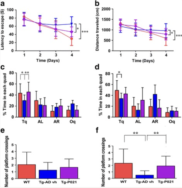
Figure 1: Changes in parameters of the Morris Water Maze test measuring improved cognitive functioning
Overall, the study found that beginning P021 treatment during the early synaptic-compensation period produced sustained protection against several features of neurodegenerative dysfunction in the 3 × Tg-AD model. The treatment restored or preserved multiple dendritic and synaptic markers, enhanced dentate gyrus neurogenesis, increased CREB activity, and improved spatial and short-term memory performance. The authors reported that approximately 18 months of treatment did not produce weight loss, tumors, or apparent signs of pain in the treated mice [1].
2) The review completed by researchers Kazim and Iqbal concluded that neurotrophic factor mimetics represent a promising strategy for addressing AD because they may target neurodegeneration through mechanisms that are complementary to approaches directed exclusively at amyloid or tau. The authors emphasized that synaptic loss correlates more closely with cognitive impairment than either amyloid plaque burden or neurofibrillary tangle burden. Consequently, restoring neurogenesis, dendritic integrity, and synaptic plasticity was presented as a potentially important therapeutic strategy. Native neurotrophic factors such as BDNF and CNTF have demonstrated neuroprotective and neurogenic properties, but their clinical development has been limited by poor pharmacokinetic characteristics, inadequate BBB penetration, short plasma half-lives, and systemic adverse effects. Small-molecule mimetics were therefore presented as an approach intended to retain beneficial neurotrophic activity while improving drug-like properties [2].
The review identified P021 as one of the most promising CNTF-derived mimetics discussed. P021 was reported to enhance adult hippocampal neurogenesis, promote neuronal and synaptic plasticity, and improve cognitive performance in several preclinical models. Its mechanism was associated primarily with inhibition of leukemia inhibitory factor (LIF) signaling and increased BDNF expression. The authors described how the compound was developed from the biologically active region of CNTF and subsequently modified to improve BBB penetration. P021 was reported to promote proliferation and differentiation of adult hippocampal progenitor cells and to increase the expression of synaptic markers. Earlier work in normal C57BL/6 mice demonstrated effects on neurogenesis, synaptic markers, and cognition, while studies in aged Fisher rats indicated that oral P021 could improve cognitive aging while enhancing neurogenesis and decreasing tau levels [2].
One of the most important findings concerned tau pathology in 3xTg-AD mice. When P021 was administered beginning at nine to ten months of age and continued for 12 months, the compound significantly reduced abnormal tau phosphorylation at both moderate and severe stages of AD-like pathology. Immunohistochemical analysis using AT8 demonstrated reduced phosphorylated tau burden in the hippocampal CA1 and subiculum regions in both the 15–16-month group receiving six months of treatment and the 21–22-month group receiving 12 months of treatment. Western blot analyses in the older animals further demonstrated significant reductions in tau phosphorylation at PHF-1-associated sites, Ser396/Ser404, and 12E8-associated sites, Ser262/Ser356. Importantly, total tau measured with a pan-tau antibody did not significantly differ between groups, suggesting that P021 primarily influenced abnormal tau phosphorylation rather than simply reducing total tau protein.
The effects of P021 on amyloid pathology were more limited than its effects on tau. The review reported a significant reduction in soluble Aβ1–40 and Aβ1–42 in the cortex of the 15–16-month animals receiving six months of P021 treatment. However, this effect was not observed in the 21–22-month animals after 12 months of treatment, and no significant effect on insoluble Aβ was identified. Histological examination showed a trend toward reduced thioflavin-S-positive plaque burden in the CA1 region of the hippocampus, but the overall effect on established plaque pathology was substantially less pronounced than the effect on tau. The authors interpreted these findings as being consistent with a reduction in Aβ generation rather than enhanced clearance. Thus, the principal disease-modifying effect of P021 appeared to involve tau phosphorylation and neuroregenerative mechanisms, with a more modest influence on amyloid-related pathology.
Another major finding was the ability of P021 to restore impaired hippocampal neurogenesis. In 15–16-month-old 3xTg-AD mice, the number of Ki-67-positive proliferating cells and DCX-positive immature neurons in the dentate gyrus was significantly reduced compared with controls. P021 treatment corrected these abnormalities, demonstrating that the compound could enhance both cellular proliferation and the generation of immature neurons during established AD-like pathology. The review further reported that P021 ameliorated deficits in MAP2, a marker associated with dendritic structure, and synaptophysin, a presynaptic marker. These improvements were detected in hippocampal regions including CA3 and CA1, respectively. The findings were interpreted as evidence that P021’s neurogenic effects were accompanied by preservation or restoration of neuronal and synaptic architecture [2].
These cellular and molecular improvements were associated with improved cognitive function. The review reported that P021 treatment rescued cognitive impairment in 3xTg-AD mice at moderate-to-severe stages of pathology. Earlier studies with the related P6 compound had demonstrated improvements in spatial reference memory and short-term episodic memory, and P021 subsequently reproduced beneficial effects on cognition while possessing improved BBB permeability and pharmacokinetic properties. The authors linked these cognitive benefits to enhanced dentate gyrus neurogenesis, improved neuronal plasticity, restoration of synaptic proteins, and reduction of pathological tau phosphorylation.
Finally, the authors proposed a mechanistic model in which P021 increases BDNF expression, activating the TrkB–PI3K–AKT signaling pathway and increasing inhibitory phosphorylation of GSK-3β at Ser9. Reduced GSK-3β activity would consequently decrease abnormal tau phosphorylation and potentially reduce amyloidogenic processing of APP. The review also emphasized that P021 possessed favorable characteristics for further drug development, including a plasma half-life greater than three hours, high stability in artificial intestinal and gastric fluids, and BBB permeability. Up to one year of administration reportedly produced no adverse effects in the 3xTg-AD or wild-type mice examined. Overall, the authors concluded that P021 represents a promising preclinical neuroregenerative and synaptic-repair strategy for AD because it simultaneously affects neurogenesis, synaptic integrity, cognition, and pathological tau [2].
Disclaimer
**LAB USE ONLY**
*This information is for educational purposes only and does not constitute medical advice. THE PRODUCTS DESCRIBED HEREIN ARE FOR RESEARCH USE ONLY. All clinical research must be conducted with oversight from the appropriate Institutional Review Board (IRB). All preclinical research must be conducted with oversight from the appropriate Institutional Animal Care and Use Committee (IACUC) following the guidelines of the Animal Welfare Act (AWA).
Citations
[1] Baazaoui N, Iqbal K. Prevention of dendritic and synaptic deficits and cognitive impairment with a neurotrophic compound. Alzheimers Res Ther. 2017;9(1):45. Published 2017 Jun 27. doi:10.1186/s13195-017-0273-7
[2] Kazim SF, Iqbal K. Neurotrophic factor small-molecule mimetics mediated neuroregeneration and synaptic repair: emerging therapeutic modality for Alzheimer’s disease. Mol Neurodegener. 2016;11(1):50. Published 2016 Jul 11. doi:10.1186/s13024-016-0119-y
Peptides Prefer the Cold
In order to reduce peptide breakdown, keep peptides refrigerated at all times but DO NOT FREEZE.
P21 is sold for laboratory research use only. Terms of sale apply. Not for human consumption, nor medical, veterinary, or household uses. Please familiarize yourself with our Terms & Conditions prior to ordering.
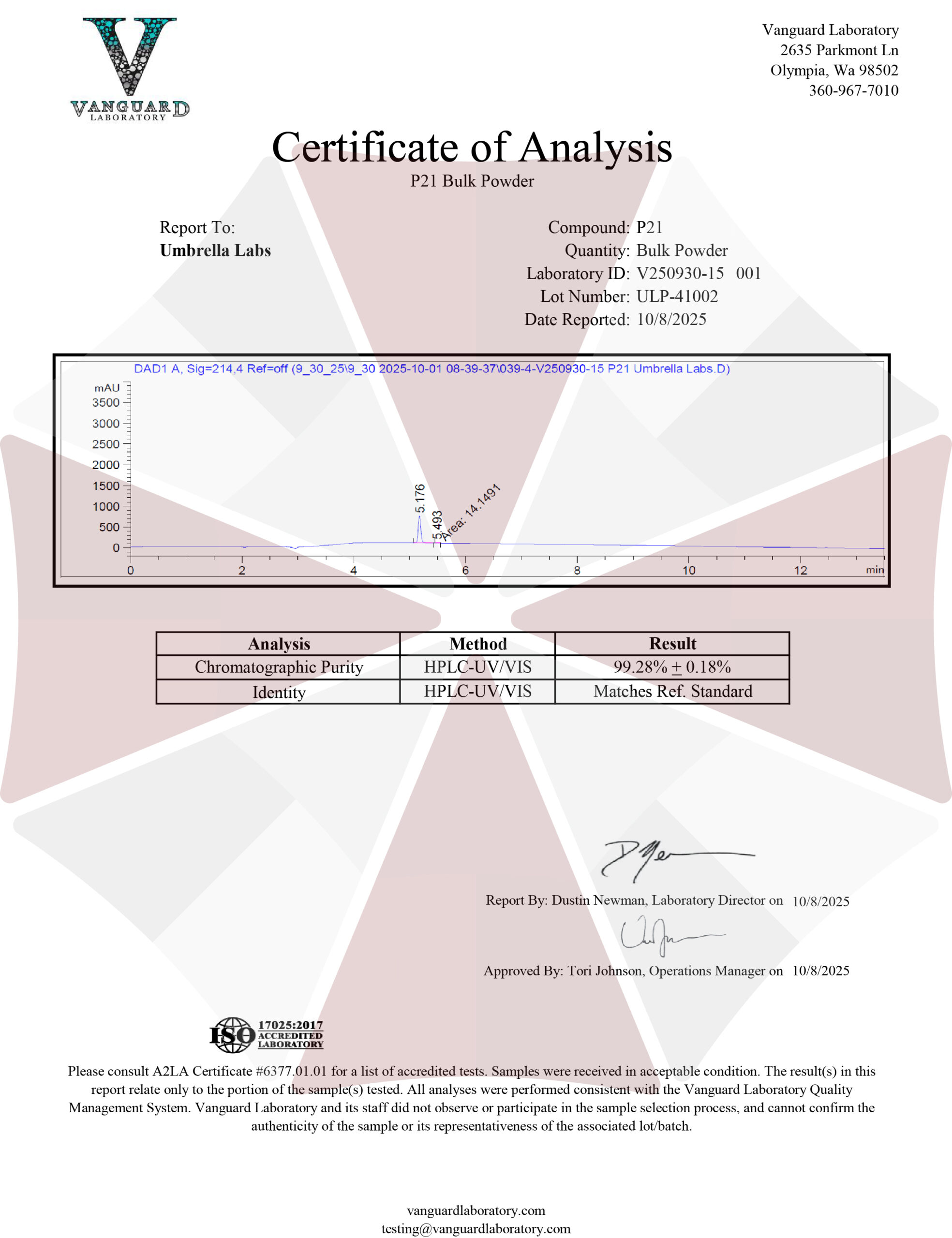
| File Name | View/Download |
| 2025-10-08-Umbrella-Labs-P21-Certificate-of-Analysis-COA.pdf |  |
VIEW CERTIFICATES OF ANALYSIS (COA)