

Description
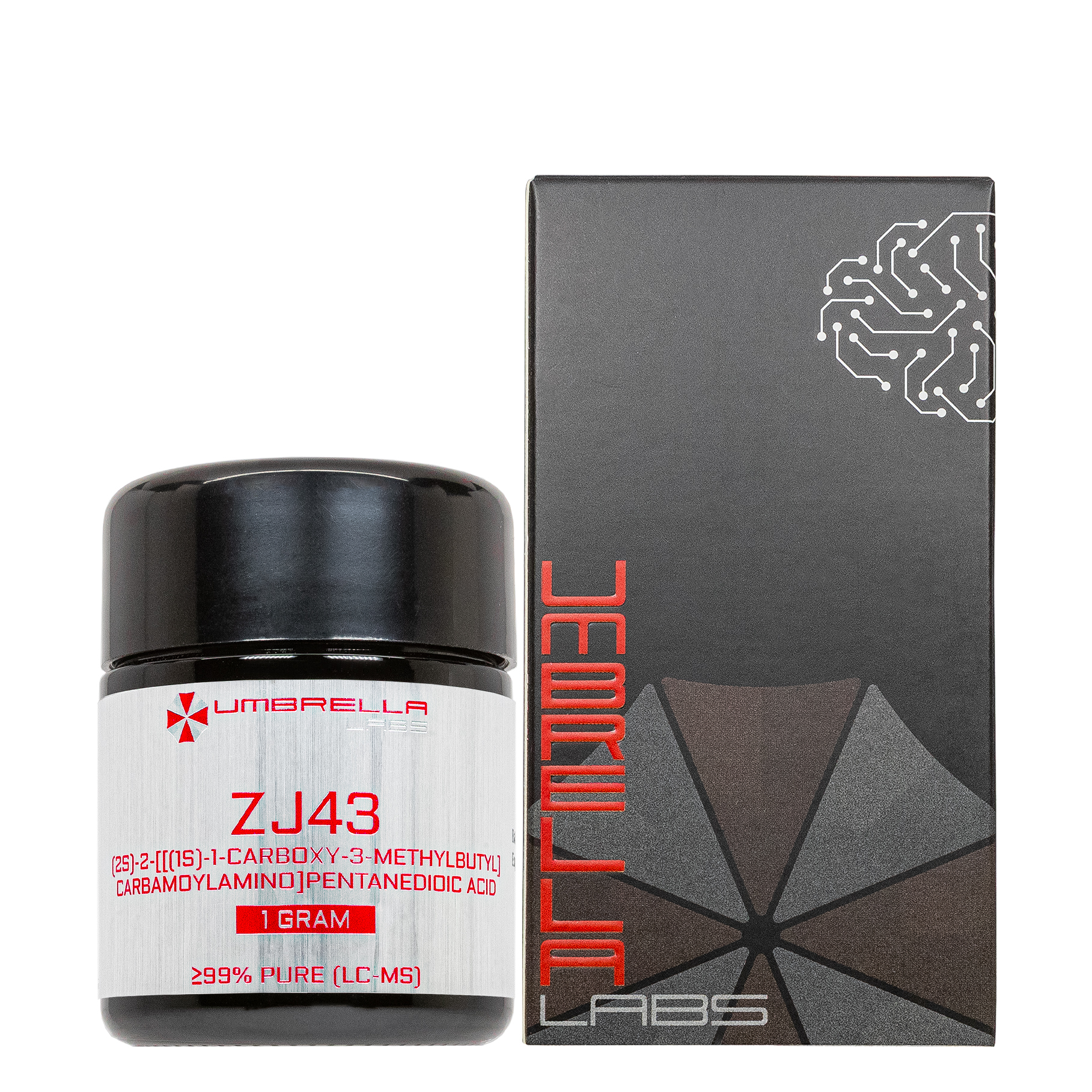
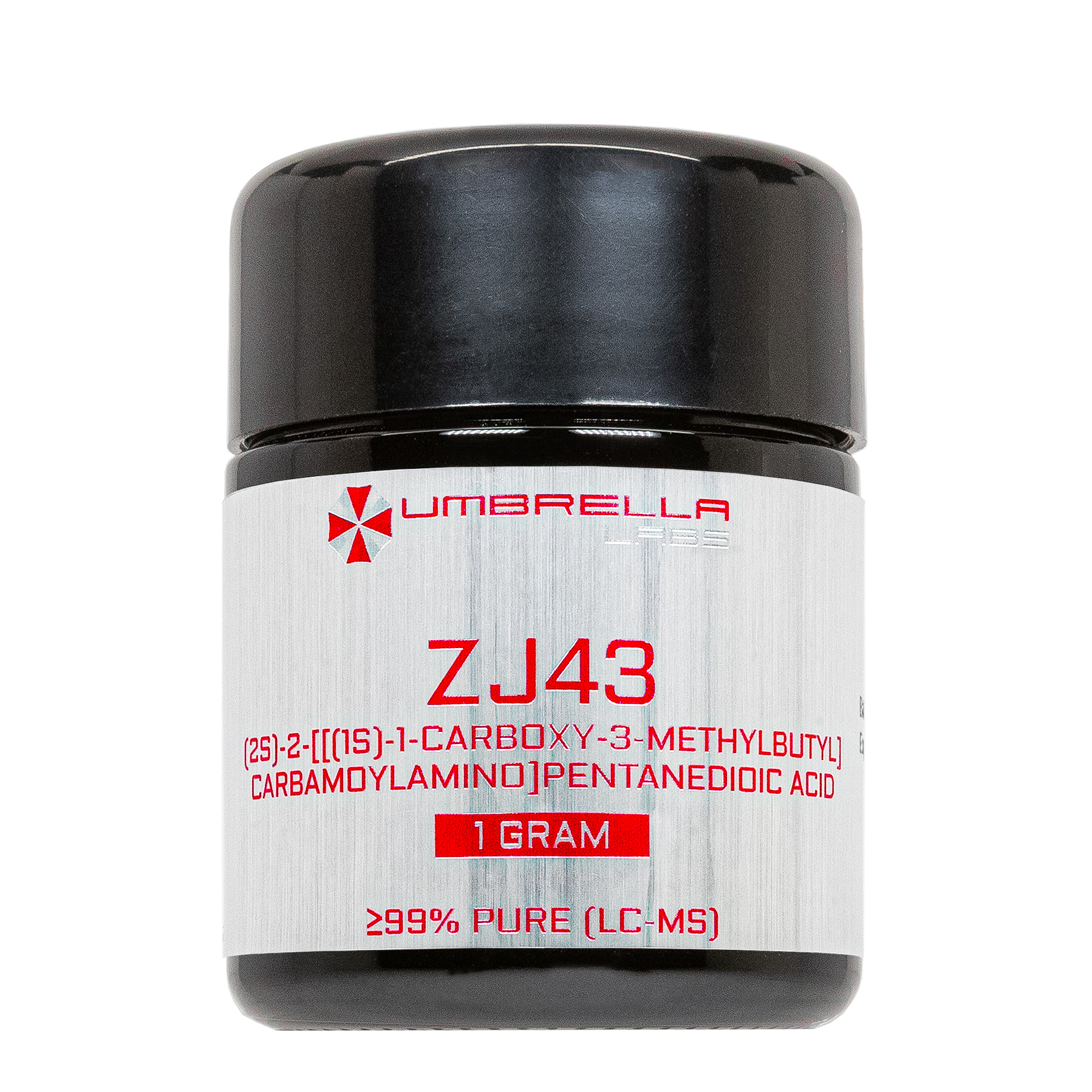
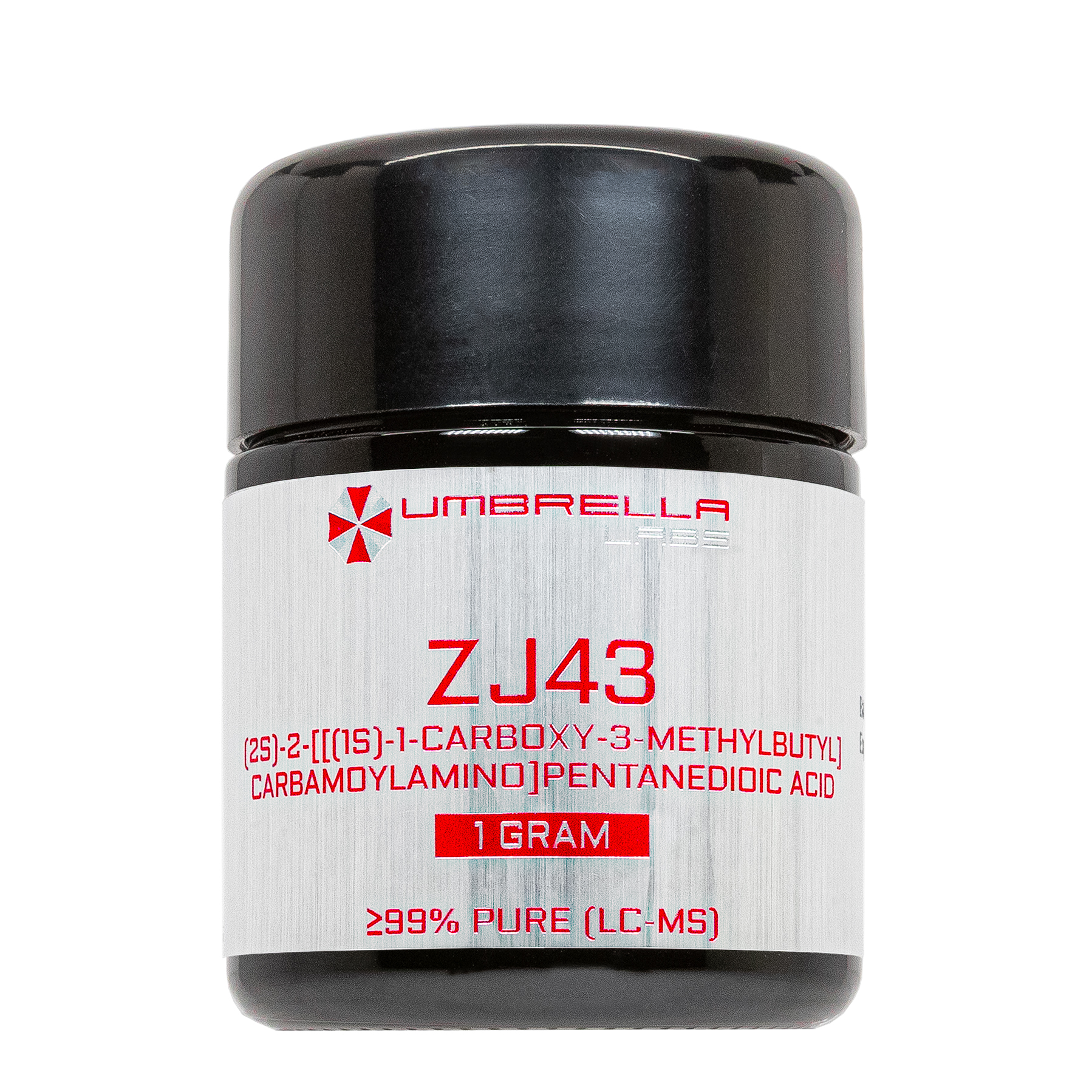
ZJ43 Nootropic Powder







| CAS Number | 723331-20-2 |
| Other Names | ZJ 43, CID 11722553, CHEMBL596351, SCHEMBL17241614, DTXSID60471136, YDB33120 |
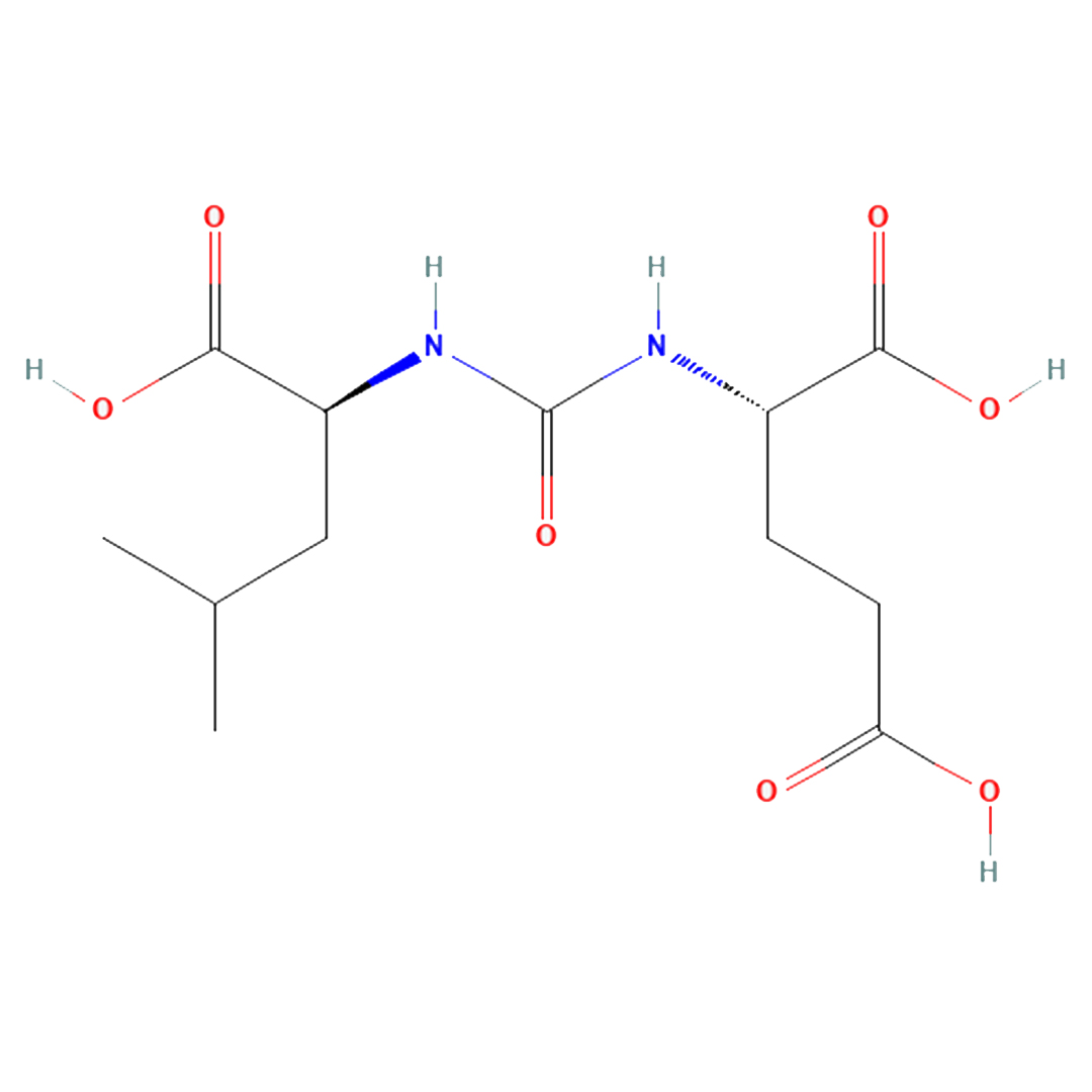
| IUPAC Name | (2S)-2-[[(1S)-1-carboxy-3-methylbutyl]carbamoylamino]pentanedioic acid |
| Molecular Formula | C₁₂H₂₀N₂O₇ |
| Molecular Weight | 304.30 |
| Purity | ≥99% Pure (LC-MS) |
| Powder Availability |  1 gram |
| Storage | Store in cool dry environment, away from direct sunlight. |
| Terms | All products are for laboratory developmental research USE ONLY. Products are not for human consumption. |
What is ZJ43?
ZJ43 is an experimental inhibitor of glutamate carboxypeptidase II (GCPII), also known as N-acetylated-α-linked acidic dipeptidase (NAALADase), that has been extensively investigated for its potential neuroprotective and cognitive-enhancing properties. By selectively inhibiting GCPII, ZJ43 prevents the breakdown of the endogenous neuropeptide N-acetylaspartylglutamate (NAAG), thereby increasing NAAG concentrations within the central nervous system. Elevated NAAG activates presynaptic metabotropic glutamate receptor 3 (mGluR3), which reduces excessive glutamate release and helps maintain excitatory neurotransmission within a physiologically balanced range. Through this mechanism, ZJ43 has demonstrated promising effects in preclinical models of traumatic brain injury, stroke, neuropathic pain, epilepsy, neurodegenerative disease, and cognitive impairment, where it has been shown to reduce excitotoxic neuronal damage, attenuate neuroinflammation, and improve functional recovery. Although ZJ43 has not advanced to clinical use, its unique mechanism of enhancing endogenous glutamatergic regulation has made it an important research compound for investigating novel therapeutic strategies aimed at preserving cognitive function and protecting the brain from glutamate-mediated injury.
Main Research Findings
1) In a phencyclidine model of schizophrenia, ZJ43 administration was shown to inhibit abnormal locomotor activity.
2) Treatment with ZJ3 was found to inhibit synaptic plasticity and transmission in the amygdala in an animal model of pain.
Selected Data
1) The study by researchers Olszewski et al was designed to evaluate the therapeutic potential of the novel glutamate carboxypeptidase (GCPII/NAALADase) inhibitor ZJ43 in an established phencyclidine (PCP)-induced animal model of schizophrenia. Because PCP produces behavioral abnormalities that resemble both the positive and negative symptoms of schizophrenia through noncompetitive antagonism of NMDA receptors, the investigators hypothesized that increasing endogenous levels of the neuropeptide N-acetylaspartylglutamate (NAAG) would activate group II metabotropic glutamate receptors (mGluRs), particularly mGluR3, thereby attenuating PCP-induced behavioral disturbances. To test this hypothesis, the researchers first synthesized and chemically characterized ZJ43, evaluated its biochemical selectivity and potency, and then examined its effects on PCP-induced locomotor activity and stereotypic behaviors in rats. In addition, pharmacological antagonism of group II mGluRs was used to determine whether any behavioral effects of ZJ43 were mediated through enhanced endogenous NAAG signaling [1].
The synthesis of ZJ43 involved a multistep organic chemistry procedure beginning with protected amino acid intermediates that were converted into a urea-based analog of NAAG. Each intermediate product was purified and structurally verified using standard analytical techniques, including proton and carbon nuclear magnetic resonance spectroscopy, optical rotation measurements, and elemental analysis. Following chemical synthesis, ZJ43 was evaluated for its ability to inhibit glutamate carboxypeptidase II (GCPII), the primary extracellular enzyme responsible for hydrolyzing NAAG after synaptic release. Human GCPII was expressed in transfected Chinese hamster ovary cells, and membrane preparations containing the enzyme were isolated for enzymatic assays. NAAG hydrolysis was quantified using a fluorescent glutamate detection assay in which glutamate liberated during enzymatic cleavage was measured with the Amplex Red Glutamic Acid Assay. Dose-response curves were generated to calculate IC50 values, and inhibitory constants were determined using the Cheng-Prusoff equation. Similar experiments were also performed using cloned glutamate carboxypeptidase III (GCPIII) to determine whether ZJ43 inhibited both known NAAG peptidases.
To determine the pharmacological specificity of ZJ43, the investigators examined whether the compound interacted directly with metabotropic glutamate receptors or NMDA receptors. Membranes expressing cloned group I, II, and III mGluRs were exposed to ZJ43, and intracellular second messenger signaling was measured to assess agonist or antagonist activity. Additional receptor-binding studies were performed through the National Institute of Mental Health Psychoactive Drug Screening Program to evaluate possible interactions with numerous neurotransmitter receptors and transporters, including dopamine, serotonin, adrenergic, opioid, GABAergic, cholinergic, cannabinoid, and histamine receptors, as well as the PCP and MK-801 binding sites of the NMDA receptor. Whole-cell patch-clamp electrophysiology was also performed in cultured rat cerebellar granule neurons to determine whether ZJ43 directly activated or inhibited NMDA receptor-mediated currents or altered spontaneous excitatory postsynaptic currents. These experiments were designed to confirm that any behavioral effects resulted from inhibition of NAAG degradation rather than direct receptor activation [1].
Behavioral experiments were conducted using adult male Sprague-Dawley rats housed under standardized laboratory conditions with unrestricted access to food and water. Animals were acclimated to the testing environment before experimentation and were used only once to eliminate habituation effects. On each testing day, rats were first placed in automated locomotor activity chambers for a 20-minute baseline recording. Animals then received intraperitoneal injections of either saline or 150 mg/kg of ZJ43, with some groups also receiving 1 mg/kg of the selective group II mGluR antagonist LY341495. Twenty minutes later, rats received either 10 mg/kg of PCP or saline before being returned to the activity chambers for an additional 160 minutes of continuous behavioral recording. Automated photobeam systems quantified locomotor activity by measuring the distance traveled throughout the observation period [1].
In addition to automated locomotor measurements, trained observers who were blinded to treatment assignments manually recorded stereotypic behaviors using a structured time-sampling protocol. One-minute behavioral observations were performed every five minutes throughout the 200-minute testing period. The investigators quantified multiple behaviors commonly associated with PCP administration, including remaining completely still, sniffing, stereotypic mouth movements, falling while walking, tremors, circling behavior, total head movements, head bobbing, and sideways head movements. Statistical analyses of locomotor activity employed generalized estimating equations to account for repeated measurements over time, while one-way analyses of variance were used to compare frequencies of individual stereotypic behaviors among treatment groups. Through this comprehensive combination of biochemical characterization, receptor pharmacology, electrophysiology, and behavioral assessment, the investigators sought to determine whether inhibition of NAAG degradation by ZJ43 could selectively reduce schizophrenia-like behaviors through activation of endogenous group II metabotropic glutamate receptor signaling rather than through direct effects on NMDA or other neurotransmitter receptors [1].
2) The study conducted by researchers Adedoyin et al investigated the role of the endogenous neuropeptide NAAG in regulating synaptic transmission and inflammatory pain within the laterocapsular division of the central nucleus of the amygdala (CeLC). Specifically, the investigators examined whether inhibition of NAAG degradation by the glutamate carboxypeptidase inhibitor ZJ43 could suppress glutamatergic neurotransmission, prevent inflammation-induced synaptic plasticity, and reduce behavioral manifestations of inflammatory pain. To accomplish this, the researchers combined behavioral pain assessments, systemic drug administration, whole-cell patch-clamp electrophysiology, pharmacological manipulation of glutamate receptors, and analysis of synaptic transmission in acute mouse brain slices. The experimental design allowed both the behavioral consequences of inflammatory pain and the underlying physiological mechanisms within a defined pain-processing pathway to be evaluated [2].
Adult male C57BL/6 mice were housed under standardized laboratory conditions with unrestricted access to food and water and maintained on a 12-hour light-dark cycle. Peripheral inflammatory pain was induced by injecting 20 μL of 5% formalin into the plantar surface of the left hind paw while mice were anesthetized with isoflurane. Control animals received equivalent saline injections. Following recovery, behavioral testing was performed at multiple time points to characterize the development of inflammatory hypersensitivity. Thermal hyperalgesia was measured using the Hargreaves plantar apparatus before injection and at 1, 3, 6, and 24 hours afterward. Mechanical allodynia was assessed using an electronic von Frey anesthesiometer before treatment and again at 1, 6, and 24 hours after injection. Withdrawal latencies and withdrawal thresholds were averaged across repeated trials for each time point to ensure measurement reliability.
To determine whether inhibition of NAAG degradation could modify the development of inflammatory pain, a separate cohort of mice received intraperitoneal injections of the NAAG peptidase inhibitor ZJ43 at a dose of 150 mg/kg. The compound was administered twice, once 30 minutes before formalin injection and again approximately 8 hours later. Based on previous pharmacokinetic observations from the investigators’ laboratory, this dosing schedule maintained enzyme inhibition during the period when inflammatory plasticity developed but allowed peptidase activity to recover before behavioral testing and electrophysiological recordings were performed 24 hours later. Mechanical withdrawal thresholds were then compared between formalin-treated animals receiving ZJ43 and untreated formalin controls to determine whether early inhibition of NAAG degradation prevented persistent pain sensitization [2].
Electrophysiological experiments were conducted using acute coronal brain slices containing the laterocapsular central nucleus of the amygdala. Brain slices were prepared from untreated mice as well as animals sacrificed 1, 6, or 24 hours after saline or formalin injection. Whole-cell patch-clamp recordings were obtained from CeLC neurons while stimulating spinoparabrachial afferent fibers that constitute a major nociceptive pathway to the amygdala. Artificial cerebrospinal fluid was continuously perfused over the tissue, and inhibitory GABAergic transmission was pharmacologically blocked to isolate excitatory glutamatergic responses. Input-output curves were generated by progressively increasing stimulus intensity and measuring the amplitude of evoked excitatory postsynaptic currents (eEPSCs). Additional current-clamp recordings evaluated intrinsic neuronal excitability by applying incremental depolarizing current injections and measuring action potential firing frequency and rheobase current. The relative contributions of NMDA and AMPA receptors were assessed by applying the selective NMDA receptor antagonist CPP and calculating NMDA/AMPA current ratios.
To investigate the physiological role of endogenous NAAG, pharmacological agents were applied directly to the brain slices during electrophysiological recordings. ZJ43 was tested across concentrations ranging from 0.01 to 100 μM to establish a dose-response relationship, after which 0.1 μM was selected for subsequent experiments because it produced approximately half-maximal inhibition of excitatory transmission. The investigators also applied purified NAAG, the selective group II metabotropic glutamate receptor agonist SLx-3095-1, and the competitive group II mGluR antagonist LY341495 to determine whether the effects of ZJ43 depended upon activation of presynaptic group II receptors. Miniature excitatory postsynaptic currents (mEPSCs) were recorded in the presence of tetrodotoxin and CPP to determine whether endogenous NAAG altered spontaneous glutamate release through presynaptic mechanisms. Additional experiments compared the effects of these pharmacological manipulations in brain slices obtained before inflammation and at both 6 and 24 hours after formalin treatment. Finally, recordings were also obtained following stimulation of cortical afferents traveling through the external capsule to determine whether inflammation-induced plasticity was specific to the spinoparabrachial pain pathway rather than representing generalized changes throughout the amygdala [2].
Discussion
1) The study performed by Olszewski et al demonstrated that ZJ43 is a highly potent and selective inhibitor of glutamate carboxypeptidase that significantly reduced several schizophrenia-like behaviors produced by PCP in rats without directly interacting with glutamate receptors or other major neurotransmitter systems. Biochemical characterization showed that ZJ43 inhibited human GCPII with an IC50 of approximately 2.4 nM and a Ki of 0.8 nM, making it one of the most potent NAAG peptidase inhibitors reported at the time. The compound also inhibited glutamate carboxypeptidase III, although with lower potency. These findings confirmed that ZJ43 effectively blocks the enzymatic degradation of NAAG, thereby increasing the availability of this endogenous peptide neurotransmitter after synaptic release.
Importantly, receptor screening experiments demonstrated that ZJ43 did not function as either an agonist or antagonist at cloned group I, II, or III metabotropic glutamate receptors despite its structural similarity to NAAG. Likewise, the compound failed to interact with NMDA receptor PCP or MK-801 binding sites or with an extensive panel of dopamine, serotonin, adrenergic, opioid, cholinergic, GABAergic, cannabinoid, histamine, and monoamine transporter targets. Whole-cell electrophysiological recordings further confirmed that ZJ43 neither activated NMDA receptor-mediated currents nor altered spontaneous NMDA-mediated excitatory postsynaptic currents, indicating that its behavioral effects resulted from inhibition of NAAG degradation rather than direct receptor modulation [1].
Behavioral testing revealed that PCP administration produced the expected robust increase in locomotor activity, characterized by dramatic elevations in total distance traveled throughout the observation period. Pretreatment with ZJ43 significantly attenuated this PCP-induced hyperlocomotion, reducing overall motor activity without producing obvious sedation when administered alone. Automated locomotor recordings demonstrated that rats receiving ZJ43 before PCP traveled significantly shorter distances than animals treated with PCP alone, indicating that increasing endogenous NAAG signaling effectively suppressed one of the principal behavioral manifestations of NMDA receptor blockade. To determine whether this protective effect required activation of group II metabotropic glutamate receptors, investigators administered the selective antagonist LY341495 before ZJ43 treatment. Blocking group II receptors completely abolished the beneficial effects of ZJ43, restoring PCP-induced locomotor activity to levels comparable with or exceeding those observed after PCP alone. Interestingly, LY341495 administered with PCP also tended to increase locomotor activity beyond that produced by PCP alone, suggesting that endogenous NAAG normally exerts a tonic inhibitory influence on PCP-induced behavioral activation through group II mGluRs even in the absence of peptidase inhibition.
The effects of ZJ43 extended beyond overall locomotor activity to several individual behaviors associated with PCP-induced stereotypy. Rats treated with PCP spent very little time remaining completely still, reflecting profound behavioral activation. Pretreatment with ZJ43 significantly increased the amount of time animals remained motionless, partially restoring normal behavioral patterns, whereas coadministration of LY341495 eliminated this improvement. Sniffing behavior increased substantially following PCP administration and remained elevated after ZJ43 treatment. Unlike other behavioral measures, ZJ43 did not reduce PCP-induced sniffing and showed a tendency to enhance this behavior slightly. Administration of LY341495 abolished both the PCP-induced and ZJ43-associated increases in sniffing, suggesting that this particular behavioral response may itself be mediated through activation of group II metabotropic glutamate receptors and may therefore respond differently to enhanced NAAG signaling than other PCP-induced behaviors [1].
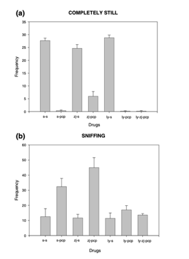
Figure 1: Changes in the frequency of the animals to A) stay still vs B) sniff, following experimental treatment
Analysis of discrete stereotypic behaviors demonstrated that ZJ43 selectively reduced several of the abnormal motor patterns induced by PCP. PCP markedly increased the frequency of falling while walking, repetitive circling, head movements, and head bobbing, all of which were significantly attenuated by pretreatment with ZJ43. Although reductions in stereotypic mouth movements, tremors, and sideways head movements did not always reach statistical significance, each displayed consistent trends toward improvement relative to PCP-treated animals. In every behavioral category showing improvement, the addition of LY341495 reversed the effects of ZJ43, with behavioral frequencies returning to levels similar to or greater than those observed following PCP treatment alone. Figures presented in the study clearly illustrated these effects, showing lower frequencies of circling, falling, and repetitive head movements in ZJ43-treated rats, whereas animals receiving both ZJ43 and the group II antagonist exhibited behavioral responses nearly indistinguishable from those of the PCP-only groups. These findings strongly supported the hypothesis that the therapeutic actions of ZJ43 depended upon enhanced activation of endogenous group II metabotropic glutamate receptors by accumulated NAAG rather than any intrinsic pharmacological activity of the inhibitor itself [1].
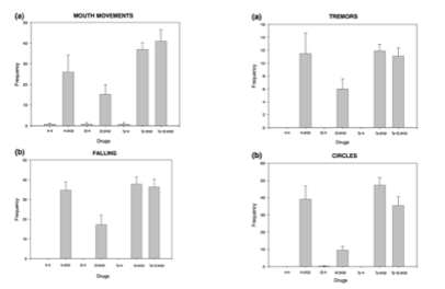
Figure 2: Changes in the frequency of abnormal motor patterns induced by PCP following experimental treatment
Overall, the investigators concluded that inhibition of NAAG peptidase represents a promising strategy for modulating glutamatergic neurotransmission in disorders characterized by NMDA receptor hypofunction. The combined biochemical and behavioral findings demonstrated that ZJ43 effectively increased endogenous NAAG signaling, selectively reduced multiple schizophrenia-like behaviors induced by PCP, and produced these effects through activation of group II metabotropic glutamate receptors rather than direct interaction with glutamate receptors or other neurotransmitter systems. Although certain behaviors, particularly sniffing, were not improved and in some cases appeared enhanced, the consistent reversal of ZJ43’s therapeutic effects by LY341495 provided compelling pharmacological evidence that endogenous NAAG functions as an important regulator of PCP-induced behavioral activation. The authors concluded that NAAG peptidase inhibitors such as ZJ43 may represent a novel therapeutic approach for treating specific behavioral components of schizophrenia by enhancing physiological glutamatergic modulation instead of directly targeting dopamine or NMDA receptor systems [1].
2) The study by Adedoyin et al demonstrated that endogenous NAAG plays an important physiological role in regulating excitatory neurotransmission within the laterocapsular central nucleus of the amygdala and that inhibition of NAAG degradation by the peptidase inhibitor ZJ43 both reduced inflammation-induced synaptic plasticity and prevented the development of persistent pain behaviors. Behavioral experiments first confirmed that intraplantar formalin injection produced a reliable model of inflammatory pain. Mice developed pronounced thermal hyperalgesia during the early stages of inflammation, with thermal withdrawal latency significantly reduced at one and three hours after formalin administration compared with both saline-treated and naive control animals [2].
By six hours, thermal withdrawal responses in all groups had largely converged because of habituation to repeated testing, and withdrawal latencies returned to baseline by twenty-four hours. Mechanical sensitivity followed a different time course. Formalin-treated mice developed significant mechanical allodynia in the injected hind paw that persisted at one, six, and twenty-four hours after injection, while the contralateral hind paw also demonstrated increased sensitivity by the twenty-four-hour time point. Pretreatment with ZJ43 before formalin administration and a second dose eight hours later prevented the development of ipsilateral mechanical allodynia measured twenty-four hours after inflammation, indicating that early inhibition of NAAG degradation interrupted the processes responsible for long-term inflammatory sensitization.
Electrophysiological recordings revealed that peripheral inflammation produced substantial increases in excitatory synaptic transmission within the CeLC. Input-output analyses showed little change one hour after formalin injection, but by six hours there was a significant increase in the amplitude of eEPSCs, indicating enhanced glutamatergic transmission. These increases became even more pronounced twenty-four hours after inflammation when recordings were performed in magnesium-free artificial cerebrospinal fluid. Despite the larger evoked currents, analysis of the NMDA-to-AMPA receptor current ratio demonstrated no significant differences between control and inflamed animals, suggesting that the enhanced synaptic responses did not result from altered postsynaptic glutamate receptor composition. Instead, current-clamp recordings demonstrated that CeLC neurons exhibited markedly increased intrinsic excitability twenty-four hours after inflammation. These neurons generated higher action potential firing frequencies in response to depolarizing current injections, while rheobase current showed a nonsignificant tendency to decrease. Together, these findings indicated that inflammatory pain produced both enhanced presynaptic glutamatergic transmission and increased postsynaptic neuronal responsiveness within this amygdala pain-processing pathway [2].
Pharmacological experiments established that endogenous NAAG normally suppresses excitatory neurotransmission through activation of presynaptic group II metabotropic glutamate receptors. Application of ZJ43 to brain slices from untreated mice produced a concentration-dependent reduction in eEPSC amplitude, reaching approximately fifty percent inhibition at the highest effective concentration. Based on these findings, the investigators selected 0.1 μM ZJ43 for subsequent experiments because it produced approximately half-maximal inhibition. This reduction in synaptic transmission was completely prevented by co-application of the selective group II mGluR antagonist LY341495, demonstrating that the inhibitory effect of ZJ43 depended upon enhanced activation of endogenous group II receptors by accumulated NAAG. Similar reductions in eEPSC amplitude were produced by direct application of purified NAAG and the selective group II receptor agonist SLx-3095-1.
In contrast, administration of LY341495 alone had no significant effect on basal synaptic transmission in control brain slices, suggesting that endogenous NAAG does not tonically activate these receptors under resting conditions. Miniature EPSC recordings further demonstrated that ZJ43 significantly reduced the frequency of spontaneous excitatory events without altering their amplitude, providing strong evidence that endogenous NAAG acts presynaptically to inhibit glutamate release rather than modifying postsynaptic receptor sensitivity [2].
The influence of endogenous NAAG changed substantially following peripheral inflammation. Six hours after formalin injection, ZJ43 continued to significantly reduce eEPSC amplitude, and this inhibitory effect remained sensitive to blockade by LY341495, indicating that NAAG-mediated regulation of glutamate release was still functional during the early stages of inflammatory plasticity. However, by twenty-four hours after inflammation the effectiveness of ZJ43 was dramatically diminished, producing only minimal reductions in excitatory transmission despite the continued ability of the selective group II agonist SLx-3095-1 to strongly suppress eEPSCs.
The investigators interpreted these findings as evidence that inflammation altered endogenous NAAG signaling rather than receptor responsiveness, possibly through decreased NAAG release or changes in the relative expression of mGluR2 and mGluR3 receptors. Importantly, systemic administration of ZJ43 around the time of formalin injection completely prevented the inflammation-induced enhancement of synaptic transmission observed twenty-four hours later. This normalization occurred specifically within the spinoparabrachial-amygdala pathway, whereas stimulation of cortical afferents traveling through the external capsule failed to demonstrate similar inflammatory potentiation, confirming that the observed synaptic plasticity was selective for pain-processing circuits [2].
Overall, the findings demonstrated that endogenous NAAG serves as an important physiological regulator of glutamatergic neurotransmission within the amygdala by activating presynaptic group II metabotropic glutamate receptors to suppress glutamate release. Peripheral inflammation progressively enhanced excitatory synaptic transmission and neuronal excitability within the CeLC while simultaneously reducing the effectiveness of endogenous NAAG-mediated inhibition. Early pharmacological inhibition of NAAG degradation with ZJ43 prevented both the development of inflammation-induced mechanical allodynia and the long-term enhancement of excitatory synaptic transmission, indicating that augmentation of endogenous NAAG signaling can interrupt the establishment of pathological pain-related plasticity. The authors concluded that NAAG peptidase inhibition represents a promising therapeutic strategy for limiting central sensitization and inflammatory pain by preserving endogenous glutamatergic regulation within specific nociceptive pathways rather than directly suppressing neuronal activity throughout the central nervous system [2].
Disclaimer
**LAB USE ONLY**
*This information is for educational purposes only and does not constitute medical advice. THE PRODUCTS DESCRIBED HEREIN ARE FOR RESEARCH USE ONLY. All clinical research must be conducted with oversight from the appropriate Institutional Review Board (IRB). All preclinical research must be conducted with oversight from the appropriate Institutional Animal
Citations
[1] Olszewski RT, Bukhari N, Zhou J, et al. NAAG peptidase inhibition reduces locomotor activity and some stereotypes in the PCP model of schizophrenia via group II mGluR. J Neurochem. 2004;89(4):876-885. doi:10.1111/j.1471-4159.2004.02358.x
[2] Adedoyin MO, Vicini S, Neale JH. Endogenous N-acetylaspartylglutamate (NAAG) inhibits synaptic plasticity/transmission in the amygdala in a mouse inflammatory pain model. Mol Pain. 2010;6:60. Published 2010 Sep 22. doi:10.1186/1744-8069-6-60
ZJ43 is sold for laboratory research use only. Terms of sale apply. Not for human consumption, nor medical, veterinary, or household uses. Please familiarize yourself with our Terms & Conditions prior to ordering.
| File Name | View/Download |
| Coming Soon! |  |
VIEW CERTIFICATES OF ANALYSIS (COA)