

Description
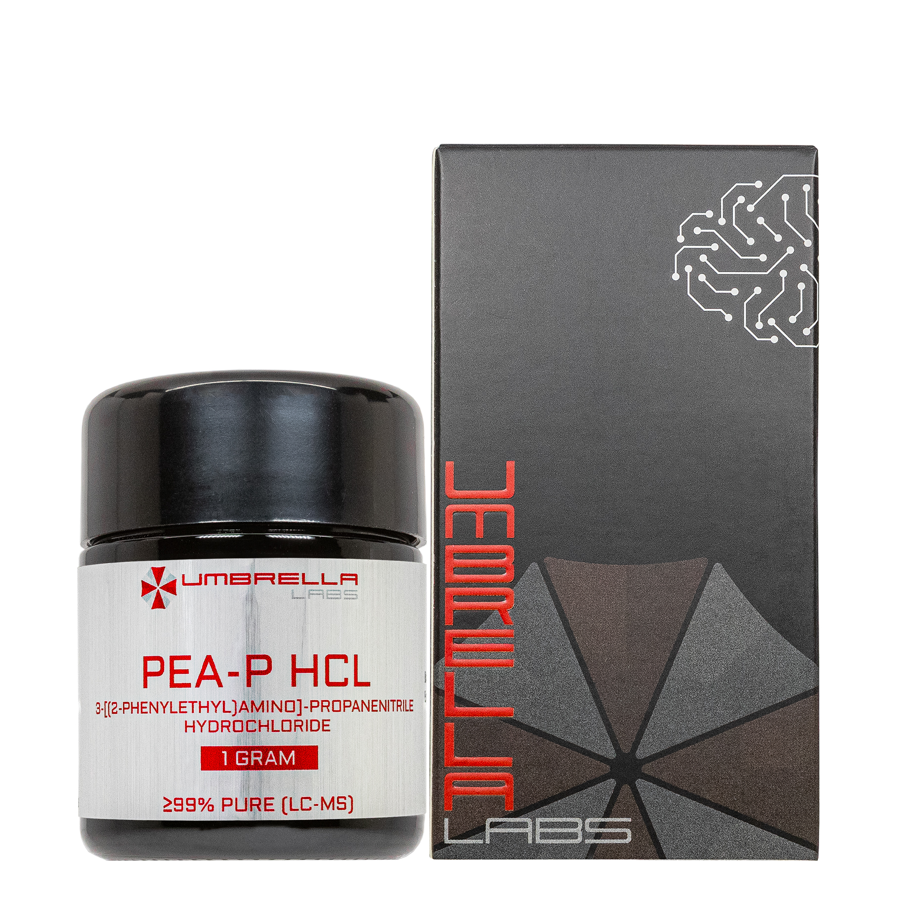
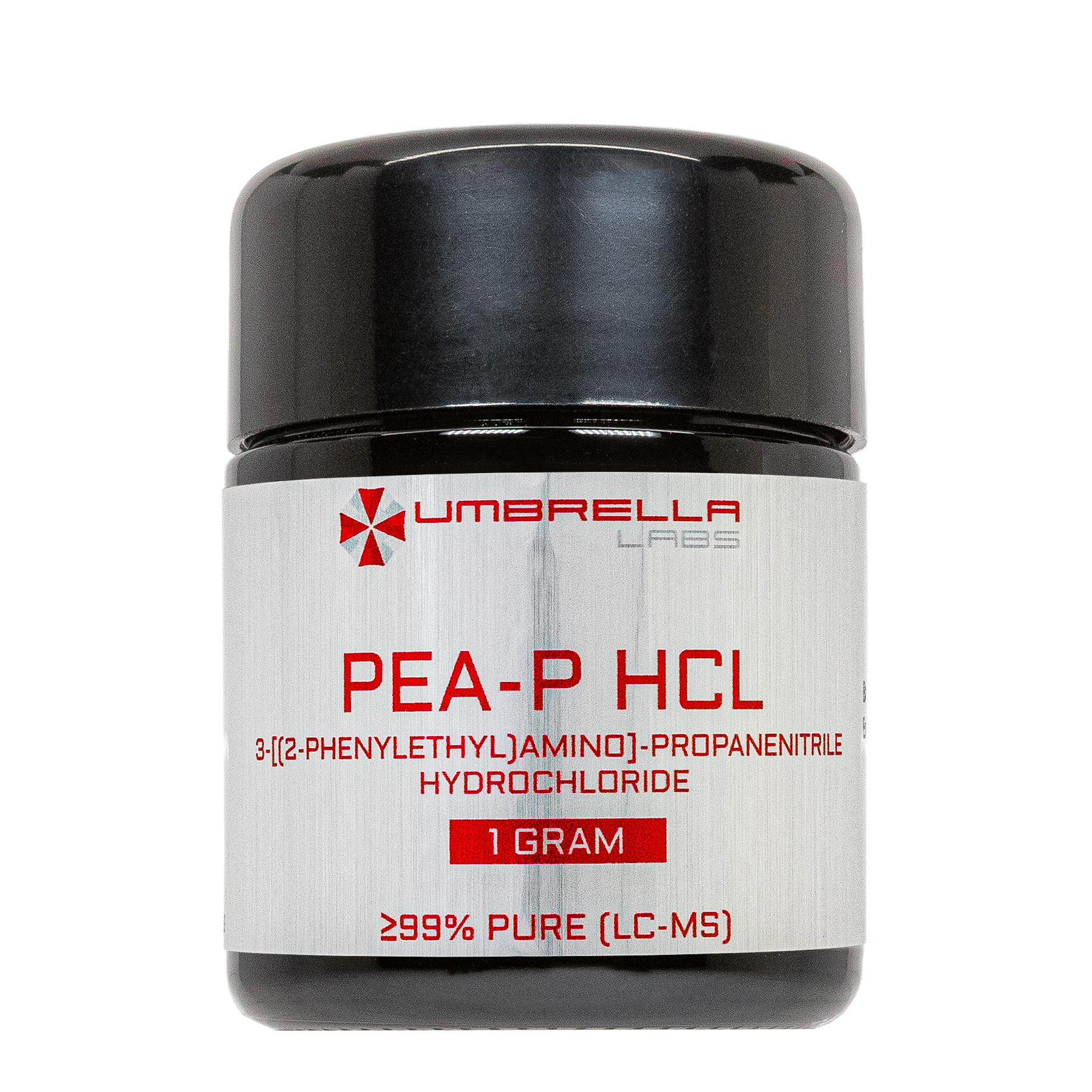
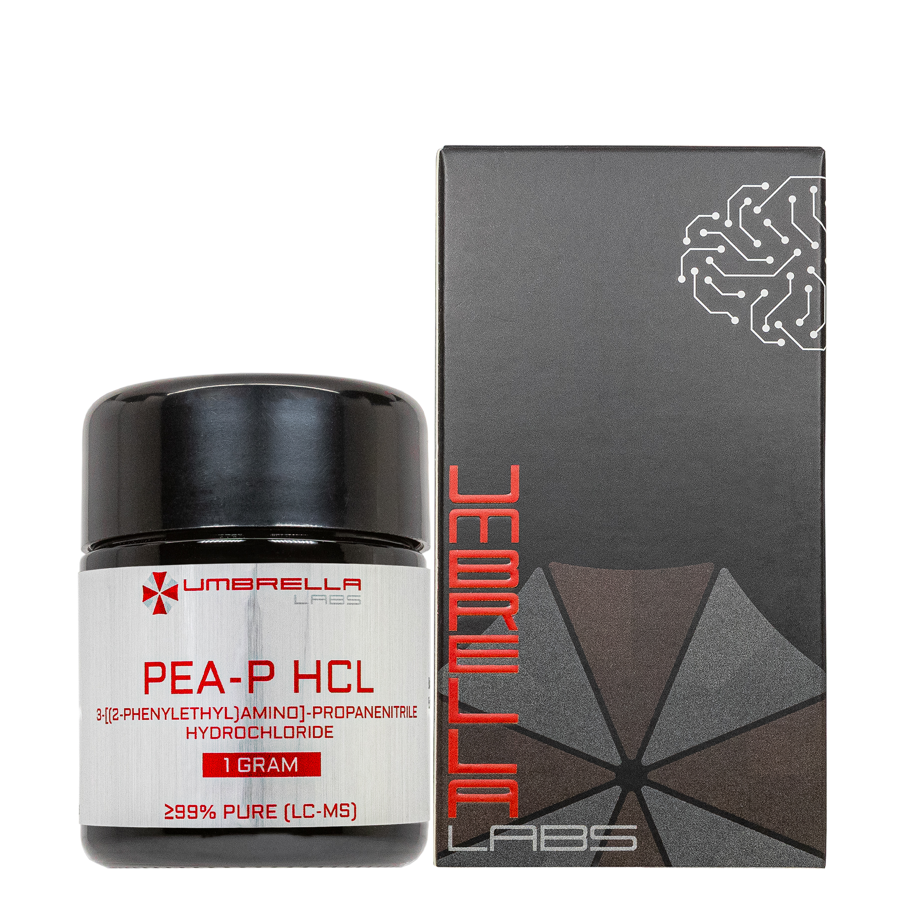
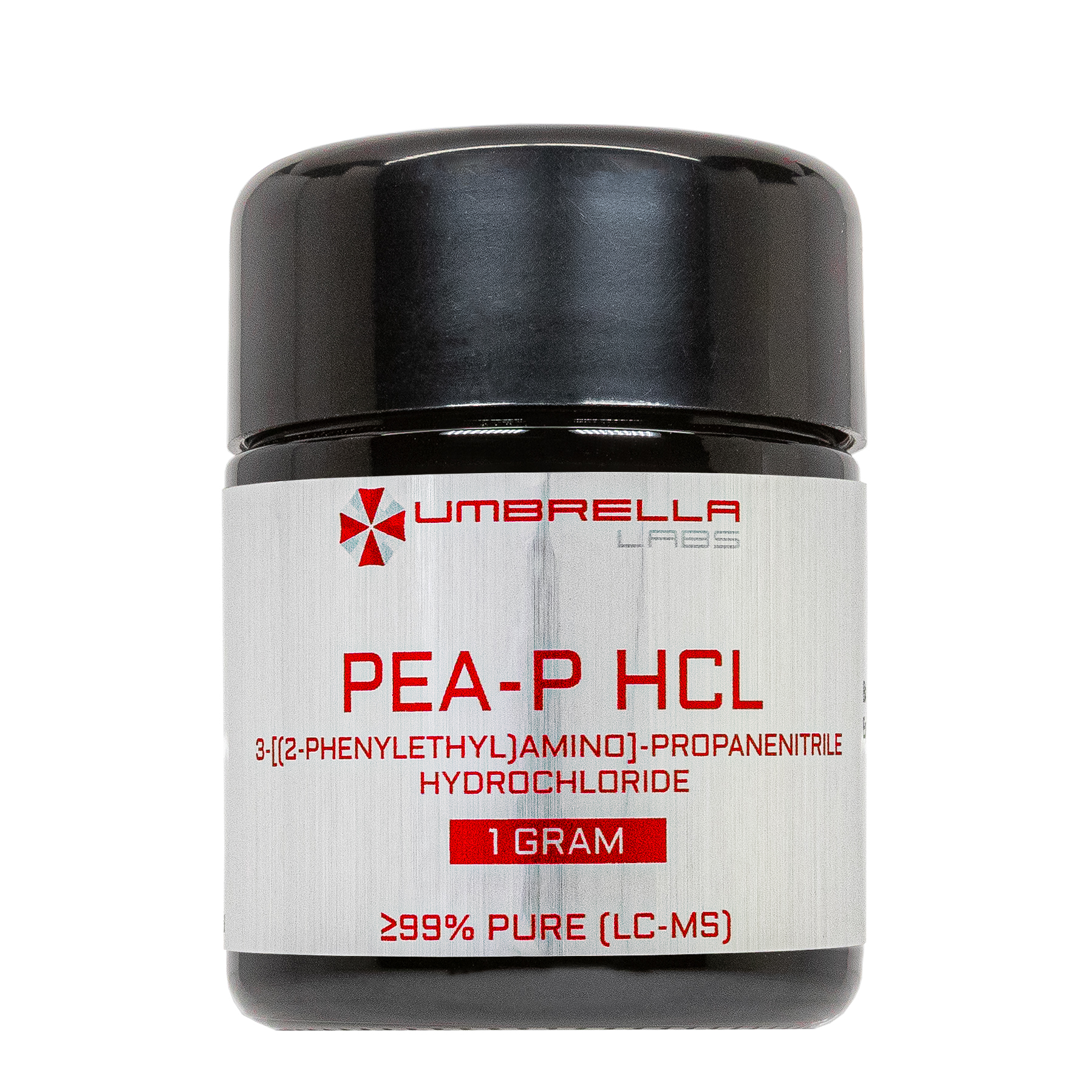
PEA-P HCl Nootropic Powder (1 Gram)







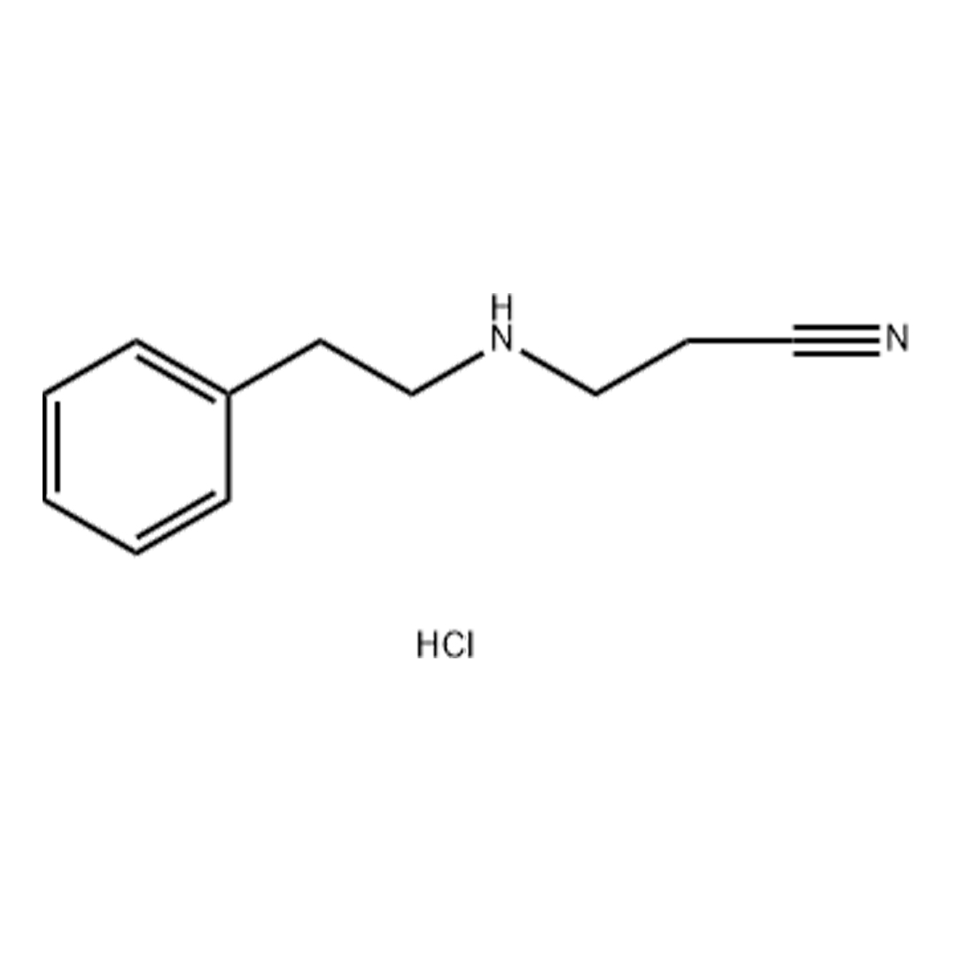
| CAS Number | 73605-24-0 |
| Other Names | 3-PHENETHYLAMINO-PROPIONITRILEHYDROCHLORIDE, PEAP |
| IUPAC Name | 3-[(2-phenylethyl)amino]-propanenitrile hydrochloride |
| Molecular Formula | C₁₁H₁₄N₂•HCl |
| Molecular Weight | 210.71 |
| Molecular Sequence | C1=CC=C(C=C1)CCNCCC#N.Cl |
| Purity | ≥99% Pure (LC-MS) |
| Liquid Availability |  30mL liquid (100mg/mL, 3000mg bottle) |
| Powder Availability | 60 capsules (50mg/capsule, 3000mg total)
|
| Storage | Store in a dry, cool, dark place. For best preservation, store at 4°C or colder away from bright light. |
| Terms | All products are for laboratory developmental research USE ONLY. Products are not for human consumption. |
What is PEA-P HCl?
PEA-P HCl (β-Phenylethylamine Priprionitrile Hydrochloride) is a synthetic formulation of the naturally occurring trace amine phenylethylamine, a compound produced in small amounts within the central nervous system that plays a role in regulating neurotransmitter activity. PEA has attracted considerable attention as a potential nootropic due to its ability to influence dopaminergic and noradrenergic signaling, pathways that are closely associated with attention, motivation, mood, and cognitive performance. The hydrochloride salt form enhances the compound’s stability for research and supplemental use. Current studies suggest that transient increases in PEA levels may promote alertness, mental energy, and emotional well-being. As a result, PEA-P HCl continues to be investigated for its potential applications in cognitive enhancement and mood support.
Main Research Findings
1) PEA-P and its derivatives were shown to have selective inhibitory effects on dopamine transportation indicating its potential to be used to treat neuropsychiatric disorders.
2) PEA-P reduces depression-like phenotypes induced by corticosterone use by activating the BDNF/TrkB/CREB signaling pathways.
Selected Data
1) The study by the research team of Kundu et al was designed to identify structural characteristics of 29 β-phenethylamine (β-PEA) derivatives that influence dopamine transporter (DAT) inhibition and to determine how these compounds interact with the human dopamine transporter at the molecular level. In addition to evaluating dopamine reuptake inhibition, the investigators performed molecular docking simulations to predict ligand-binding interactions and conducted functional experiments to determine how selected β-PEA derivatives affected dopamine D2 receptor (D2R) signaling. Together, these complementary biochemical, computational, and cellular approaches allowed the researchers to establish structure–activity relationships (SAR) and investigate the functional consequences of DAT inhibition [1].
The experimental studies utilized human embryonic kidney (HEK-293) cells that were either stably or transiently transfected with human dopamine transporter (hDAT) or dopamine D2 receptor constructs. Cells were maintained under standard culture conditions in minimal essential medium supplemented with fetal bovine serum, penicillin, and streptomycin. The test compounds consisted of 29 structurally distinct β-PEA derivatives. GBR12909, a well-characterized dopamine reuptake inhibitor, served as the reference compound during dopamine uptake experiments, while radiolabeled dopamine ([³H]-dopamine) and radiolabeled sulpiride ([³H]-sulpiride) were used to quantify dopamine transport and D2 receptor surface expression, respectively.
To measure dopamine transporter inhibition, investigators employed a dopamine reuptake assay using HEK-293 cells expressing human DAT. Cells grown in 24-well plates were first washed with uptake buffer containing physiological concentrations of sodium, potassium, calcium, magnesium, glucose, and antioxidants. The cells were then incubated with individual β-PEA derivatives for approximately 20 minutes at 37°C before the addition of radiolabeled dopamine. Uptake was allowed to proceed for five minutes, after which the reaction was terminated by washing the cells with ice-cold buffer. Cells were subsequently lysed overnight using sodium dodecyl sulfate, and the accumulated radioactivity was quantified with a liquid scintillation counter. Compounds that inhibited dopamine uptake by at least 20–30% during the initial screening were selected for detailed concentration-response testing. Dose-response curves were generated for these compounds, and half-maximal inhibitory concentration (IC₅₀) values were calculated using GraphPad Prism software to compare their inhibitory potency [1].
To better understand the molecular basis of dopamine transporter inhibition, the researchers performed computational docking simulations using generated structural models of the human dopamine transporter. The three-dimensional transporter structure underwent energy minimization before docking analyses were conducted. Two compounds demonstrating particularly strong dopamine reuptake inhibition, compounds 9 and 28, were selected for detailed modeling. Because both molecules possess chiral centers, all possible stereoisomers were generated and evaluated individually. Docking simulations were carried out using four different scoring algorithms to improve confidence in predicted ligand orientations and binding affinities. The docking search space was centered around the known dopamine-binding pocket, and predicted ligand interactions with amino acid residues within the transporter were visualized using molecular graphics software. This computational approach enabled comparison of stereochemical preferences, hydrogen bonding interactions, hydrophobic contacts, and predicted binding stability among the various isomers.
To determine whether inhibition of dopamine reuptake influenced dopamine receptor function, the investigators conducted a dopamine D2 receptor endocytosis assay. HEK-293 cells were transiently transfected with dopamine D2 receptors together with either a mock vector or human DAT. Following transfection and cell plating, cultures were stimulated with dopamine to induce receptor internalization. Surface D2 receptor expression was subsequently quantified using radiolabeled sulpiride, a hydrophilic ligand that selectively binds receptors remaining on the plasma membrane. Some experimental groups were pretreated with selected β-PEA derivatives prior to dopamine exposure to determine whether DAT inhibition altered receptor trafficking. The amount of surface receptor remaining after agonist stimulation served as an indirect measure of receptor endocytosis and allowed the investigators to evaluate whether dopamine transporter activity modulated dopamine receptor signaling [1].
The collected data were expressed as mean ± standard deviation, and statistical analyses were performed using one-way analysis of variance followed by Tukey’s post hoc multiple comparison test. Differences were considered statistically significant when the probability value was less than 0.05. By integrating radiolabeled uptake assays, molecular docking simulations, structure–activity relationship analyses, and functional receptor trafficking experiments, the investigators established a comprehensive framework for evaluating how structural modifications of β-phenethylamine derivatives influence dopamine transporter inhibition and downstream dopaminergic signaling [1].
2) The study conducted by the research team of Lee et al employed both in vitro and in vivo experimental models to investigate the antidepressant effects of 2-phenylethylamine (PEA) and to determine whether its therapeutic actions were mediated through the brain-derived neurotrophic factor (BDNF)/tropomyosin receptor kinase B (TrkB)/cAMP response element-binding protein (CREB) signaling pathway. The investigators combined cellular, molecular, and behavioral techniques to evaluate the effects of PEA on hippocampal neuronal morphology, synaptic protein expression, neurotrophic signaling, and depression-like behaviors induced by chronic corticosterone (CORT) exposure. Male C57BL/6N mice were housed under controlled environmental conditions with unrestricted access to food and water and maintained on a standard 12-hour light/dark cycle throughout the experiments [2].
To examine the cellular effects of PEA, primary hippocampal neurons were isolated from embryonic day 18 (E18) mouse embryos and cultured under standard conditions. Neurons were plated on poly-D-lysine- and laminin-coated coverslips and maintained in neurobasal medium supplemented with serum, B27, and N2. At ten days in vitro, neurons were transfected with a green fluorescent protein (GFP) plasmid using Lipofectamine to enable visualization of dendritic spines. Four days after transfection, cultured neurons were exposed to corticosterone at a concentration of 50 μM for 24 hours to induce cellular changes associated with depression, followed by treatment with PEA at a concentration of 100 μM for an additional 24 hours. Confocal microscopy was subsequently used to examine dendritic spine density and morphology. An investigator blinded to treatment groups quantified dendritic spines located on secondary dendrites, and individual spines were classified into filopodia, thin, mushroom, and stubby subtypes according to established morphological criteria [2].
To determine whether PEA influenced synaptic integrity and neurotrophic signaling, cultured hippocampal neurons underwent biochemical analysis following treatment. Total cellular protein was isolated using radioimmunoprecipitation assay (RIPA) buffer, and protein concentrations were measured before separation by SDS-polyacrylamide gel electrophoresis and transfer to polyvinylidene fluoride membranes for Western blot analysis. Primary antibodies against synaptophysin (SYP), postsynaptic density protein 95 (PSD95), BDNF, TrkB, CREB, phosphorylated CREB (p-CREB), and β-actin were used to evaluate changes in excitatory synaptic proteins and intracellular signaling molecules. Protein bands were visualized using chemiluminescence and quantified with ImageJ software after normalization to β-actin. Reverse transcription polymerase chain reaction (RT-PCR) was also performed to quantify mRNA expression of BDNF and TrkB using RNA extracted from cultured neurons, with glyceraldehyde-3-phosphate dehydrogenase (GAPDH) serving as the internal reference gene.
The in vivo component of the study utilized a chronic corticosterone-induced mouse model of depression. Mice received daily intraperitoneal injections of corticosterone at a dose of 20 mg/kg or saline for 21 consecutive days. Beginning on day 17, subsets of corticosterone-treated animals additionally received either 50 mg/kg of PEA or the 10 mg/kg of tricyclic antidepressant imipramine, which served as a positive control, during the final four days of corticosterone exposure. Following treatment, hippocampal tissue was collected for molecular analyses, including RT-PCR and Western blotting, to measure mRNA and protein expression of BDNF, TrkB, synaptophysin, and PSD95. These analyses were performed to determine whether the neuroprotective effects observed in cultured neurons also occurred within the hippocampus of animals exhibiting depression-like behaviors.
Behavioral testing was conducted after completion of the treatment protocol to evaluate depression- and anxiety-related behaviors. Anhedonia was assessed using the sucrose preference test, in which mice were acclimated to sucrose solution before undergoing food and water deprivation and subsequent measurement of sucrose versus water consumption. Behavioral despair was evaluated using both the tail suspension test and the forced swimming test. During the tail suspension test, mice were suspended by the tail for five minutes while immobility time was recorded. In the forced swimming test, mice were placed in a water-filled cylinder for five minutes, and durations of immobility, swimming, and climbing behaviors were quantified [2].
Anxiety-like behavior was examined using the elevated plus maze, which consisted of two open and two enclosed arms elevated above the floor. Time spent in the open and closed arms was recorded using automated video tracking software. All behavioral assessments were performed using standardized protocols commonly employed in preclinical models of depression and anxiety.By integrating neuronal culture experiments, chronic animal models, behavioral assessments, gene expression analyses, protein quantification, and morphometric evaluation of dendritic spines, the investigators comprehensively examined the mechanisms through which PEA influences depression-related neuroplasticity and behavioral outcomes [2].
Discussion
1) The study by researchers Kundu et al established a clear structure–activity relationship among 29 β-PEA derivatives by demonstrating that relatively small structural modifications substantially altered their ability to inhibit dopamine reuptake through the human DAT. Initial screening identified several compounds with measurable inhibitory activity, after which the most active molecules underwent concentration-response testing to determine their IC₅₀. The results showed considerable variability in potency across the different chemical classes, indicating that both aromatic substitutions and modifications to the amine-containing side chain strongly influenced DAT inhibition [1].
Among the arylalkylamine derivatives, compounds containing unsubstituted phenyl or thiophenyl groups generally exhibited stronger dopamine reuptake inhibition than compounds containing methoxy-substituted aromatic rings. In particular, methoxy substitutions markedly reduced or completely eliminated inhibitory activity, demonstrating that the electronic and steric properties of the aromatic ring are major determinants of transporter binding. Likewise, alterations in the alkylamine substituent influenced activity, with certain secondary amine substitutions producing greater inhibition than others depending on the accompanying aromatic structure. These findings demonstrated that optimal dopamine transporter inhibition requires a specific combination of aromatic and amine substitutions rather than modification of a single structural feature.
The second group of compounds, consisting of 2-(alkyl amino)-1-arylalkan-1-one derivatives, revealed additional structural requirements for effective DAT inhibition. The investigators found that compounds containing smaller heterocyclic rings, particularly pyrrolidine derivatives, consistently exhibited greater potency than molecules containing larger ring systems such as azepane or piperazine. Furthermore, extending the carbon chain attached to the amino group progressively increased inhibitory activity, with hexyl-substituted derivatives demonstrating substantially greater potency than shorter ethyl- or propyl-containing analogs. Replacing a phenyl ring with a naphthyl group also enhanced inhibitory effects in selected compounds. Among all molecules examined, compound 28, an alkyl 2-phenyl-2-(piperidin-2-yl)acetate derivative, exhibited the strongest dopamine reuptake inhibition with an IC₅₀ of approximately 68 nM, making it the most potent DAT inhibitor identified in the study. Compound 9 also demonstrated high potency among the arylalkylamine derivatives, whereas many methoxy-substituted analogs displayed little or no measurable activity. Collectively, these observations enabled the investigators to establish detailed structure–activity relationships identifying the molecular features that favor dopamine transporter inhibition [1].
Computational docking simulations further explained the experimental findings by demonstrating favorable molecular interactions between the most potent compounds and the human dopamine transporter. All four docking algorithms consistently predicted that the (S)-enantiomer of compound 9 exhibited greater binding stability than its corresponding (R)-form, while the (R,S)-stereoisomer of compound 28 produced the most favorable docking scores among its possible stereoisomers. The predicted binding poses showed that both compounds occupied the dopamine-binding pocket of DAT in a manner similar to endogenous dopamine. Hydrogen bonding with the conserved Asp79 residue, together with multiple hydrophobic interactions involving residues located on transmembrane helices 1, 3, 6, and 8, appeared to stabilize ligand binding. Compound 28 formed additional hydrophobic contacts with Phe320, providing a structural explanation for its superior inhibitory potency compared with other derivatives. These computational results closely paralleled the biochemical assays and demonstrated that stereochemistry and specific protein-ligand interactions are major contributors to DAT affinity [1].
Functional experiments examining dopamine D2 receptor signaling demonstrated that dopamine transporter activity significantly influenced receptor regulation. Dopamine stimulation induced approximately 40% internalization of D2 receptors in cells expressing the receptor alone. However, co-expression of the dopamine transporter markedly reduced receptor endocytosis, consistent with enhanced dopamine clearance from the extracellular environment and reduced receptor activation. Pretreatment with the potent DAT inhibitors, particularly compound 28 and compound 29, significantly reversed this transporter-mediated suppression of receptor internalization by restoring extracellular dopamine availability. These findings indicate that inhibition of dopamine reuptake not only elevates synaptic dopamine concentrations but also alters downstream dopamine receptor signaling by facilitating D2 receptor activation and endocytosis. Interestingly, although compound 28 exhibited greater potency as a DAT inhibitor than compound 29, it produced a somewhat weaker restoration of D2 receptor endocytosis, suggesting that mechanisms beyond simple transporter inhibition may contribute to receptor regulation.
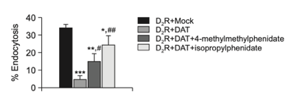
Figure 1: Disinhibitory effects of compounds 28 and 29 DAT-mediated inhibition of dopamine receptor endocytosis.
Overall, the study successfully identified multiple structural determinants governing dopamine transporter inhibition by β-phenethylamine derivatives and demonstrated that these compounds exert meaningful functional effects on dopaminergic signaling. The combined biochemical, computational, and cellular findings showed that optimal DAT inhibition depends on precise aromatic substitutions, favorable alkylamine configurations, appropriate stereochemistry, and stable molecular interactions within the transporter binding pocket. In addition to defining the structural features associated with high DAT affinity, the study demonstrated that potent dopamine reuptake inhibitors can modify dopamine D2 receptor signaling by increasing extracellular dopamine concentrations. These findings provide valuable insight into the design of selective dopamine transporter inhibitors and suggest that appropriately optimized β-phenethylamine derivatives may serve as useful lead compounds for the development of future therapeutics targeting neuropsychiatric disorders involving dopaminergic dysfunction, while also highlighting their potential psychoactive properties and the need for careful pharmacological evaluation [1].
2) The study by Lee et al demonstrated that PEA effectively reversed both the cellular and behavioral abnormalities produced by chronic CORT exposure, supporting its potential antidepressant activity through modulation of the BDNF/TrkB/CREB signaling pathway. In cultured hippocampal neurons, CORT treatment significantly impaired dendritic spine architecture, producing an overall reduction in dendritic spine density and substantial alterations in spine morphology. Specifically, CORT exposure decreased the total number of dendritic spines by approximately 37% relative to untreated controls. Morphological analysis further revealed marked reductions in mature thin and mushroom spines, accompanied by increases in immature filopodia and stubby spines, indicating impaired synaptic maturation and neuronal plasticity. Treatment with PEA largely reversed these structural abnormalities by restoring dendritic spine density and normalizing the distribution of spine subtypes, demonstrating that PEA protects hippocampal neurons from CORT-induced synaptic damage. These findings suggest that PEA promotes structural plasticity within hippocampal neurons, an effect that is considered essential for normal learning, memory, and emotional regulation [2].
PEA also restored excitatory synaptic function by reversing the reductions in synaptic proteins produced by chronic corticosterone exposure. Western blot analysis demonstrated that CORT significantly decreased expression of the presynaptic protein SYP and the postsynaptic scaffold protein PSD95, indicating substantial impairment of excitatory synapses. Following PEA treatment, expression of both proteins increased toward control levels, suggesting recovery of synaptic integrity. Additional molecular analyses showed that CORT markedly suppressed both BDNF and TrkB protein expression in cultured hippocampal neurons, while significantly reducing phosphorylation of CREB without altering total CREB expression. Administration of PEA restored BDNF and TrkB protein levels and substantially increased CREB phosphorylation, indicating reactivation of the BDNF/TrkB/CREB signaling pathway. Because this signaling cascade regulates neuronal survival, synaptic plasticity, and activity-dependent gene transcription, restoration of these molecular markers provides mechanistic evidence explaining the neuroprotective effects of PEA observed at the structural level [2].
The molecular findings obtained in cultured neurons were reproduced in the chronic CORT-treated mouse model. Long-term corticosterone administration significantly reduced hippocampal mRNA and protein expression of BDNF and TrkB, confirming disruption of neurotrophic signaling associated with depression-like pathology. Likewise, expression of the synaptic proteins SYP and PSD95 was markedly decreased at both the transcriptional and protein levels. Treatment with either PEA or the reference antidepressant imipramine significantly restored BDNF and TrkB gene expression as well as protein abundance within the hippocampus. Similar improvements were observed for SYP and PSD95, indicating recovery of excitatory synaptic function following PEA administration. The comparable effects produced by PEA and imipramine suggest that PEA possesses antidepressant-like properties capable of reversing corticosterone-induced molecular deficits that are characteristic of chronic stress and depressive disorders.
Behavioral testing demonstrated that the molecular improvements induced by PEA translated into meaningful functional recovery. In the sucrose preference test, chronic corticosterone exposure reduced sucrose consumption by approximately 44%, reflecting the development of anhedonia, a core symptom of major depressive disorder. PEA administration restored sucrose preference to levels comparable with untreated control animals, indicating recovery of normal reward-related behavior. Similarly, corticosterone significantly increased immobility during the tail suspension test, demonstrating behavioral despair. Treatment with PEA markedly reduced immobility time, producing improvements comparable to those observed with imipramine. Comparable antidepressant-like effects were observed during the forced swimming test, where corticosterone substantially increased immobility while reducing active swimming behavior. Administration of PEA significantly decreased immobility and increased swimming time, indicating improved behavioral coping responses and reduced depression-like behavior. These findings consistently demonstrated that PEA alleviated multiple behavioral manifestations of chronic stress-induced depression [2].
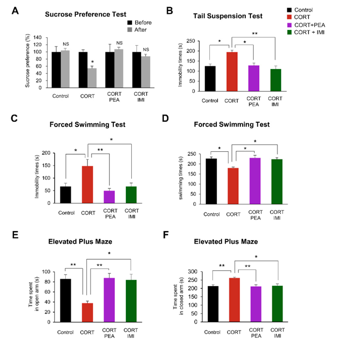
Figure 2: Changes in behavioral testing outcomes following treatment with experimental compounds following exposure to corticosterone.
PEA also improved anxiety-related behaviors produced by chronic corticosterone administration. During the elevated plus maze, CORT-treated mice spent significantly less time exploring the open arms and correspondingly more time in the enclosed arms, reflecting increased anxiety-like behavior. Treatment with PEA reversed these behavioral abnormalities by increasing time spent in the open arms while reducing time spent in the closed arms, indicating reduced anxiety. The magnitude of these behavioral improvements closely paralleled those observed following treatment with imipramine, further supporting the therapeutic potential of PEA. Collectively, the cellular, molecular, and behavioral findings demonstrated that PEA effectively counteracted corticosterone-induced depression-like pathology by restoring dendritic spine architecture, increasing excitatory synaptic protein expression, reactivating the BDNF/TrkB/CREB signaling pathway, and improving both depressive- and anxiety-like behaviors. These results provide strong evidence that enhancement of hippocampal neuroplasticity represents a primary mechanism through which PEA exerts its antidepressant effects and support further investigation of PEA as a potential therapeutic agent for stress-related mood disorders [2].
Disclaimer
**LAB USE ONLY**
*This information is for educational purposes only and does not constitute medical advice. THE PRODUCTS DESCRIBED HEREIN ARE FOR RESEARCH USE ONLY. All clinical research must be conducted with oversight from the appropriate Institutional Review Board (IRB). All preclinical research must be conducted with oversight from the appropriate Institutional Animal Care and Use Committee (IACUC) following the guidelines of the Animal Welfare Act (AWA).
Citations
[1] Kundu D, Zhu A, Kim E, et al. Potential Functional Role of Phenethylamine Derivatives in Inhibiting Dopamine Reuptake: Structure-Activity Relationship. Biomol Ther (Seoul). 2023;31(1):108-115. doi:10.4062/biomolther.2022.047
[2] Lee YJ, Kim HR, Lee CY, et al. 2-Phenylethylamine (PEA) Ameliorates Corticosterone-Induced Depression-Like Phenotype via the BDNF/TrkB/CREB Signaling Pathway. Int J Mol Sci. 2020;21(23):9103. Published 2020 Nov 30. doi:10.3390/ijms21239103
PEA-P HCl is sold for laboratory research use only. Terms of sale apply. Not for human consumption, nor medical, veterinary, or household uses. Please familiarize yourself with our Terms & Conditions prior to ordering.
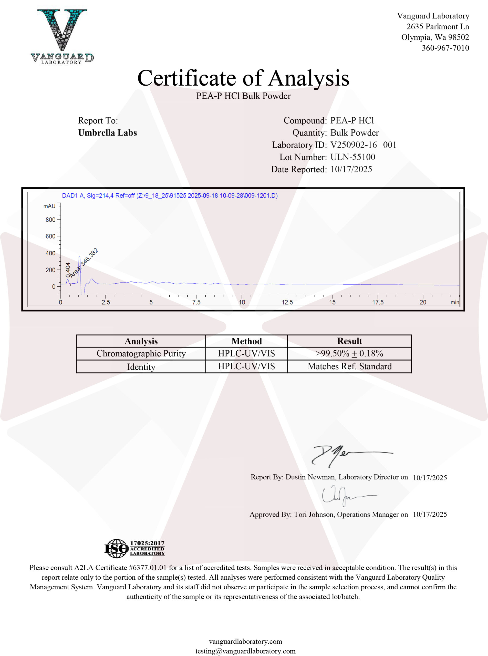
| File Name | View/Download |
| 2025-10-17-Umbrella-Labs-PEA-P-HCl-Certificate-of-Analysis-COA.pdf |  |